SPIROMETRY
SPIROMETRY. Spirometry is the measurement of the flow and volume of air entering and leaving the lungs. Static lung volumes and capacities. Tidal Volume TV The volume of air inhaled & exhaled at each breath during normal quiet breathing Inspiratory Reserve Volume IRV

SPIROMETRY
E N D
Presentation Transcript
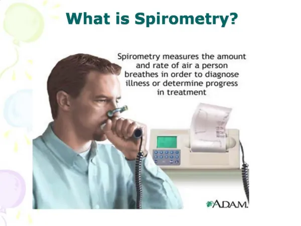
Spirometry is the measurement of the flow and volume of air entering and leaving the lungs
Static lung volumes and capacities Tidal Volume TV The volume of air inhaled & exhaled at each breath during normal quiet breathing Inspiratory Reserve Volume IRV The volume of air that can be forcefully inspired following a normal quiet inspiration Expiratory Reserve Volume ERV The volume of air that can be forcefully expired after a normal or resting expiration
Vital Capacity VC The maximum amount of air that can be exhaled after the fullest inspiration possible (TV + ERV + IRV) Inspiratory CapacityIC The maximum amount of air that can be inhaled after a normal exhalation (TV + IRV)
Residual Volume RV: The volume of air remaining in the lungs after a forceful expiration. Total Lung Capacity TLC : The total volume of the lungs (VC + RV) Functional Residual capacity FRC: The amount of air remaining in the lungs after a normal quiet expiration (ERV + RV)
Lung volumes & capacities depend on: • Age • Body size (height & weight) • Gender • Pulmonary health • Altitude • Irritants
Dynamic lung volumes: • Valuable in following the progress of a patient with chronic lung disease. • Assessing the results of a treatment • Epidemiological surveys to assess industrial hazards or to document the incidence of disease in the community • NOT a key factor in making a definitive diagnosis
Forced Vital Capacity FVC: The total volume expired by a forced maximal expiraion from a position of maximal inhalation Forced Expiratory Volume in 1 sec FEV1.0: The volume of air expired in the first second of maximal forced expiration from a position of full Inspiration
Other volumes & capacities Maximal Ventilatory Volume (MVV): maximal amount of air that a person can breathe in or out in a short period of time, typically 10, 12, or 15 secs.
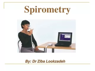
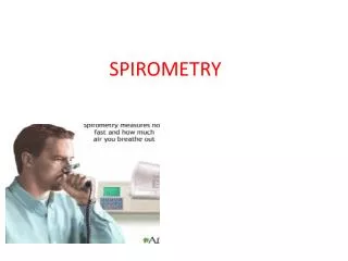
Spirometry • the measurement of the flow and volume of air entering and leaving the lungs • test of pulmonary function (PFT) • Indicator of health status or disease • Exercise fitnes
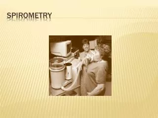
Spirometer •Apparatus used to measure static & dynamic lung volumes/capacities using a closed system. •Registers the amount and rate of air moved into or out of the lungs
2 main types: 1. Volume: records the amount of air exhaled or inhaled within a certain time* 2. Flow: measures how fast the air flows in or out as the volume of air inhaled or exhaled increases
Respiratory Diseases •Bronchitis • Asthma • COPD • Emphysema • Influenza
Obstructive Airways Disease For obstruction, review the FEV1/FVC ratio first. If this is less than Lower Limit of predicted, think obstruction, and then observe the FEV1 to determine degree ofobstruction. When FEV1/FVC is below the Lower Limit of Normal (LLN)……. Mild : FEV1 above LLN (>70% predicted) Moderate : FEV1 40-60% predicted Severe : FEV1 < 40% predicted Please check current guidelines for asthma and COPD as these may change, depending on source.
Obstructive (Bronchial Asthma) •Abnormally large TLC, but expiration ends prematurely. • Early airway closure brought about by the increased smooth muscle tone of the bronchii (asthma) or loss of radial traction from surrounding parenchyma (emphysema). • Other causes include edema of bronchial walls or secretions in the airway.
Restrictive Lung Disease: For restriction, review the FEV1/FVC ratio first. If this is normal, think restriction, and then observe FVC to determine degree of restriction. When FEV1/FVC is normal Mild : FVC below LLN (>60% predicted) Moderate : FVC 50-60% predicted Severe : FVC < 50% predicted
Restrictive Lung Disease Affects the lung tissue The inability of the lungs to expand. The lungs are small because ofFibrosis or scarring Full inflation is not possible (disease of lung lining, chest wall or abdomen. Inspiratory respiratory muscles are weak
Compliance • the ease with which the chest wall and lungs expand, degree of stiffness. • Direct effect on the amount of pressure required to increase of decrease lung volume • Low compliance = ‘stiffer’ lungs ∴more effort required to inflate alveoli • Pulmonary fibrosis → restrictive lung disease
Resistance • Degree of ease in which air can pass through the Airways • Determined by the number, length and diameter of the airways. • High resistance → individual may not be able to exhale fully ∴ some air trapped in lungs
Restrictive (Pulmonary Fibrosis): • Loss of pulmonary and chest compliance, or weakness of the respiratory muscles