Spirometry workshop
Spirometry workshop. Jo Riley Lead Respiratory Nurse. History. Borelli, 1679 – first measured volume of air that a man can inhale in a single breath

Spirometry workshop
E N D
Presentation Transcript
Spirometry workshop Jo Riley Lead Respiratory Nurse
History • Borelli, 1679 – first measured volume of air that a man can inhale in a single breath • Hutchinson,1846 – defined vital capacity as the “greatest expiration following the deepest inspiration”, designed spirometer to measure this but not accepted and died in obscurity! • Rohrer, 1915 – looked at relationship between respiratory muscle force and rate of airflow. • Peabody, 1915 – examined the relationship between vitaal capacity and breathlessness. The most common tests that we perform today are based on these early works.
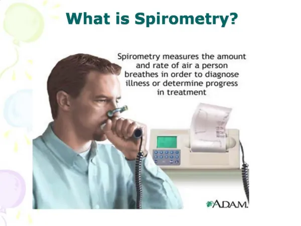
What are we measuring? • Lung volumes • Resistance to airflow In the lung function laboratory we can also assess gas transfer and residual volumes.
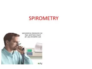
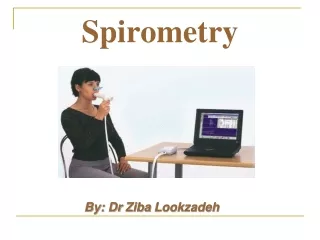
What tests can we perform outside the laboratory? • Peak flow • Spirometry
Peak flow • Measures how quickly air can be expelled from the airways • Measured within the first 100milliseconds (10th of a second) • Index of resistance to flow through the larger airways, bronchi and larger bronchioles Individuals with bronchial hypersensitivity are subject to reflex bronchoconstriction in these airways. Peak flow is an effort dependant test.
Reduced peak flow Upper airway obstruction e.g. Bronchial Ca, goitre. Obstructive airways disease e.g.asthma, COPD Advanced restrictive lung disease e.g.UIP, FA Chest wall abnormalities e.g. scoliosis, neuromuscular disease Normal peak flow Normal airways! Well controlled or stable asthma Mild COPD and asymptomatic smokers Early restrictive lung disease (Can sometimes be raised) Interpretation of peak flow
Why do we do peak flows? • For diagnostic purposes • Comparison to normal/predicted values • Reversibility testing – bronchodilator and corticosteroid • Peak flow diaries – diurnal variation and serial peak flows • Exercise challenge tests • To monitor control of asthma • Prn monitoring
Why do spirometry? • More informative than peak flow • To detect presence or absence of lung disease where there is a history or pulmonary symptoms • To confirm findings of other investigations e.g.chest x-ray or blood gasses • To establish extent of lung impairment in respiratory disease and monitor progression e.g.COPD / Fibrosis • To investigate impact of other diseases on lung function e.g. cardiac disease or neuromuscular disease • Occupational / environmental monitoring e.g. smokers, dust, asbestos • To determine effects of an intervention e.g.bronchodilator reversibility tests
Spirometry cannot - • Define the full extent of the disease e.g. In COPD many systemic as well as pulmonary effects • Define the response to therapy • Define the extent of disability that the patient experiences
Guidelines Avoid • Smoking for 24 hours • Alcohol for 4 hours • Vigorous exercise for 30 minutes • Tight clothing • Food for 2 hours • Query Using Inhalers
Check History • MI / CVA • Recent operations • Spontaneous pneumothorax • Aneurysm • Uncontrolled hypertension, angina • Ear infection • Pregnancy
Patient preparation • Record patients date of birth, height, ethnic origin • Note if the patient is currently unwell or has had a recent exacerbation • Ensure the patient is comfortable • Sit the patient in a chair with arms • Explain the purpose of the test • You may need to demonstrate the correct technique • Allow the patient practice attempts
Patient preparation To withhold or not to withhold medication? If you are doing reversibility testing: • No short acting bronchodilators for 4 hours • No long acting bronchodilators for 12 hours • No sustained release oral bronchodilators for 24 hours For routine monitoring of COPD patients: • Take all medication as usual
Inspiratory reserve vol Inspiratory capacity Vital capacity Tidal vol Expiratory reserve vol Residual vol Lung volume terminology
Terminology VC – Vital capacity, the total amount of air that acn be expelled from the lungs from full inspiration to full expiration FVC – Forced vital capacity, should be the same volume as VC but is sometimes reduced due to air trapping in COPD FEV1 – Forced expiratory volume in one second from full inspiration FEV1/FVC or FEV1% or FEV1/FVC ratio –The percentage of the FVC that is produced in the first second FEV1/VC - The percentage of the VC that is produced in the first second
Measuring vital capacity (VC) The VC is a non vorced measurement. It is often measured at the start of a session. • Patient breathes in as deeply as is comfortable • Seals lips around mouthpiece • Breathes out steadily at a comfortable pace • Continue until expiration complete • May need a nose clip • Repeat
Measuring FEV1 and FVC • Ask the patient to take a deep breath in – full inspiration • Patient to blow out forcibly, as hard and fast as possible, until there is nothing left to dispell • Encourage patient to keep blowing • For some COPD patients this can take up to 15 seconds! • Spirometer may bleep to say manoeuvre complete • Repeat the procedure twice or until reproducible results
Maintaining accuracy The most common reason for inacurate results is patient technique Common problems include: • Inadaquate or incomplete inhalation • Additional breath taken during manoeuvre • Lips not sealed around mouthpiece • A slow start to the forced exhalation • Some exhalation through the nose • Coughing
Interpreation of results • Take the best of the 3 consistent readings of FEV1 and of FVC • Find the predicted normals for your patient – Your machine may do this for you! • Get out your calculators!!
Depends on ; Age Sex Height Race Predicted Normals
Based on large population surveyse.g.ERS93, ECCS83 Predicted values are the mean values obtained from the survey No surveys conducted in elderly populations Predicted Normal values
Normal ventilatory function • FVC 80 – 120% of predicted • FEV1 80 – 120% of predicted • FEV1/FVC ratio >70%
To calculate % predicted Actual Measurement x100 Predicted Value • e.g. Actual FEV1 = 4.0 litres Predicted FEV1 = 4.0 litres 4 x 100 = 100% 4
To calculate the ratio of FEV1 to FVC (FEV1%, FEV1/FVC or FER) Actual FEV1 x100 Actual FVC e.g. FEV1 = 3.0 litres FVC = 4.0 litres 3 x100 =75% 4
Results clasification • Normal • Obstructive • Restrictive • Combined
Flow volume trace Peak expiratory flow Flow (l/second) FVC Volume (litres)
Flow volume trace Peak expiratory flow Flow (l/second) FVC Volume (litres)
Obstructive defects • COPD • Asthma • Bronchial carcinoma • Bronchiectasis
Obstructive flow volume trace Peak expiratory flow Flow (l/second) Volume (litres)
Fibrosing lung disease (CFA, UIP, EAA, rheumatiod) Parenchymal tumours Pneumoconiosis (coal workers, asbestosis, silicosis, siderosis) Byssinosis Pulmonary oedema Restrictive defectsPulmonary causes
Predicted normal curve Flow (l/second) Volume (litres) Restrictive flow volume trace
Combined defects • Severe COPD • Advanced bronchiectasis • Cystic fibrosis
Combined flow volume trace Peak expiratory flow Predicted normal curve Flow (l/second) FVC FVC Volume (litres)
?Reversibility testing??? NICE 2004 R12 In most patients suspected of having COPD, routine spirometric reversibility testing is not necessary as a part of the diagnostic process or to plan initial therapy with bronchodilators or corticosteroids. It may be misleading or unhelpful because • Repeated FEV1 measurements can show spontaneous fluctuations • Results on different occasions may be inconsistant and not reproducible • Unless change in FEV1 is >400mls a single test may be misleading • Definition of magnitude of significant change is purely arbitrary • Response to long term therapy not predicted by acute reversibility testing History is Key in distinguishing asthma from COPD
Patient preparation To withhold or not to withhold medication? If you are doing reversibility testing: • No short acting bronchodilators for 4 hours • No long acting bronchodilators for 12 hours • No sustained release oral bronchodilators for 24 hours For routine monitoring of COPD patients: • Take all medication as usual
History Suggests COPD and FEV1 < 80% predicted FEV1/FVC < 70% Diagnose COPD And follow COPD guidelines If no doubt If in doubt If still In doubt If FEV1 improves < 400 mls Bronchodilator Reversibility 400 mcg Salbutamol or equivalent Terbutaline Steroid reversibility Oral Prednisolone 30 mg daily for 2 weeks If FEV1 improves < 400 mls If FEV1 improves > 400 mls Asthma likely to be present If FEV1 improves > 400 mls