Spirometry ...
Spirometry ... . … all you ever needed to know!. How to do it . ... a quick refresher. Before you start. Prior to testing, the patient's condition should be stable (i.e. at least 6 weeks since the last exacerbation).


Spirometry ...
E N D
Presentation Transcript
Spirometry... … all you ever needed to know!
How to do it ... ... a quick refresher
Before you start • Prior to testing, the patient's condition should be stable (i.e. at least 6 weeks since the last exacerbation). • Sitting is safer than standing when performing the tests – ideally in a chair with arms and with the patient’s feet flat on the floor.
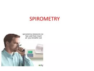
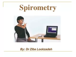
Ask the patient to ... • Breathe in maximally • Hold mouthpiece between teeth, then apply lips for an airtight seal • Hold mouthpiece between teeth, then apply lips for an airtight seal • Breath out as hard and fast as possible. The patient should aim for maximal flow at the moment expiration starts • Keep breathing out until the lungs are 'empty'
Relaxed or forced VC ... or both? • For most accuracy record relaxed as well as forced manoeuvres. Following a maximal inspiration ask the patient just to empty their lungs in a slow and sustained manner until they can blow out no more. In obstructive diseases the relaxed VC may often be far greater then the FVC. • Limit the total number of attempts to eight for each manoeuvre (forced VC, relaxed VC – and PEF if measured separately)
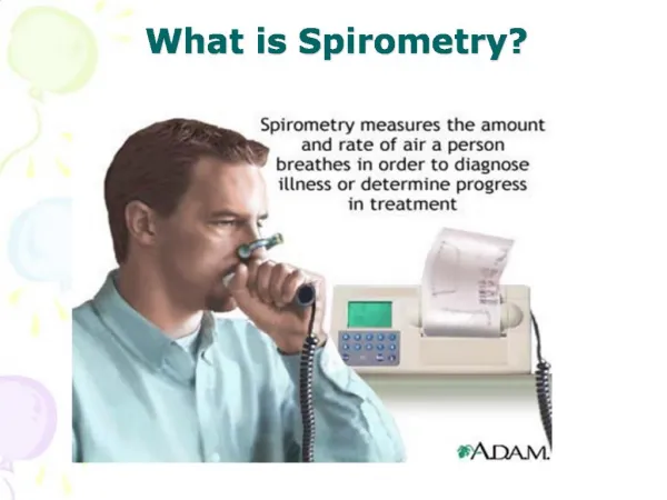
What am I measuring ... and why?
Most important measurements • PEF: Peak expiratory flow • FEV1: Forced expiratory volume measured at 1 second • VC: Vital capacity – this may forced (FVC) or relaxed (RVC – usually referred to as VC) • FEV1/VC ratio: (FEV1/FVC) x 100Percentage of FVC expelled in the first second of a forced expiration.
What does it show …? PEF • PEF is an index of resistance to airflow through the larger airways, bronchi and larger bronchioles • i.e it is a measure of how quickly air can be expelled from the airways! • Indicates presence of airflow obstruction • NB: PEFdoes not distinguish between obstruction and restriction of airflow, and may seriously underestimate the degree of airflow obstruction in COPD.
FEV1/FVC ratio • (FEV1/FVC)x100 • Percentage of FVC expelled in the first second of a forced expiration • NB: for increased accuracy use whichever vital capacity measurement (relaxed or forced) gives the best result - (FEV1/FVC)x100 or (FEV1/VC)x100
FEV1 • FEV1 demonstrates the amount of air that can be expelled forcefully in one second • This is a function of the size and elastic properties of the lungs, the calibre of the bronchial tree and the collapsibility of the airway walls • Indicates type of respiratory disease (restrictive or obstructive) • Severity of airflow limitation
Remember ... • FEV1 is reproducible and objective with well defined normal ranges • Variation on different occasions on the same patient is low (<170 ml) • Serial measurements provide evidence of disease progression
VC • VC demonstrates the volume of air that can be expired from a full inspiration • Simple measure of the size of the lungs • May be reduced due to small lung size or loss of elasticity and airway collapsedue to airflow obstruction (i.e. in restrictiveor obstructive disorders)
FEV1/FVC ratio • Useful to differentiate between obstructive and restrictive results • The hallmark of an obstructive defect is slowing of expiratory flow, so that a low proportion of the FVC is expired in the first second and the FEV1/FVC ratio is reduced • A ratio of <70% implies obstructive disease.
Obstructive or restrictive? • In the elderly, the FEV1/FVC may fall to <70% in the absence of airways obstruction, so compare to predicted values to determine if obstruction present • If the value is >70%, obstruction is effectively excluded in everyone • If the patient has a restrictive ventilatory defect the FEV1 and FVC are both reduced, but in proportion, so the FEV1/FVC ratio remains normal (greater than 75%)
Reporting results • Minimum of 3 acceptable spirometric manoeuvres • 2 highest values must be within 5% or 100 mls for FEV1 , VC and FVC • 2 highest values must be within 60 L/sec for PEF • Select highest FEV1 and VC value • Even if they are not from the same attempt
‘Best test’ results • Automatic computerised spirometers often select ‘best test’ results • ie the acceptable manoeuvre with largest value when FEV1 and VC added together • BEWARE: Spirometer does not always select highest value obtained out of all attempts !
Observe patient performing tests: • patient did not inspire to TLC/blow out to RV • a leak at the mouth/nose • obstructed mouthpiece due to tongue/teeth • poorly co-ordinated start to manoeuvre • cough within first second of blow • early termination of blow • test conducted with submaximal effort
Reference Values • Common practice to compare measurement/calculated value with a reference or predicted value • Comparison provides basis for interpretation • Not a good diagnostic tool, but may be useful to exclude disease
Interpret with caution! The use of reference values raises some concerns: • Insufficient values for certain race and age groups (e.g. >70 years)
Are the results normal? • Normal subject can expire 50-60% VC in 0.5 seconds, 75-85% VC in 1 second, 94% in 2 seconds, 97% in 3 seconds and entire VC in 4 seconds • Slightly lower ratios are nomal in elderly adults
Subjects with obstructive disease will show a reduced FEV1/VC ratio • Ratio less than 70% indicative of airflow obstruction • Subjects with restrictive disease will show a normal or supranormal FEV1/VC ratio • Flow rates minimally effected • FEV1 and VC reduced in same proportion
Remember … • Validity of interpretation depends on recognition of subject effort and co-operation performing tests • Poor effort on VC can lead to overestimation of FEV1/VC ratio • Poor effort on FEV1 can lead to misdiagnosis of disease
Deterioration over time? • Expected decrease in FEV1 and VC due to aging is approximately 25-30 ml/year
Clinically significant change • Week to week change in VC >11% • Week to week change in FEV1 >12% • Yearly changes in FEV1 or VC >15%
What do you need to know? • Is the FEV1 normal or reduced? • Is the VC normal or reduced? • Is the FEV1/VC ratio normal, reduced or high? • Is PEF normal or reduced? • Are the results within normal limits?
Do the results demonstrate an obstructive or restrictive defect? • Is this mild, moderate or severe? • Do you need to check for reversible airflow obstruction? • Which inhaler will you use? • What dose?
Normal spirometry: • FEV1, FVC, VC, PEF >80% predicted • FEV1/VC ratio >70% predicted Obstructive defect: • low FEV1 (<80%) • low FEV1/VC ratio (<70%) • VC normal or low Severity graded on % predicted for FEV1
Restrictive defect: • FEV1 normal or low • normal or high FEV1/VC ratio • low VC Severity graded on % predicted for VC Severity: Mild: 50 - 80% predicted Moderate: 30 - 49% predicted Severe: <30% predicted Reversibility: > 400 ml increase in FEV1