Spirometry Interpretation
Spirometry Interpretation. Debbie Terry- Respiratory Team Leader. Aims and Objectives. Aim of the session To gain an advanced understanding of spirometry interpretation Objectives Gain understanding of spirometry parameters Understand required standards for performing spirometry

Spirometry Interpretation
E N D
Presentation Transcript
Spirometry Interpretation Debbie Terry- Respiratory Team Leader
Aims and Objectives Aim of the session To gain an advanced understanding of spirometry interpretation Objectives • Gain understanding of spirometry parameters • Understand required standards for performing spirometry • Identify obstructive and restrictive patterns • Use spirometry tracing to help diagnose obstructive/restrictive disorders • Understand NICE guidance in relation to determining COPD severity
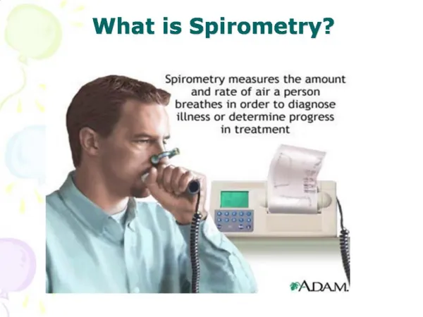
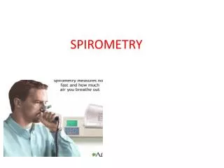
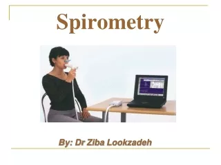
What is spirometry? • Conventionally, a spirometer is a device used to measure timed expired and inspired volumes of air. • From these measurements we can calculate how effectively and how quickly the lungs can be emptied and filled
Objectives of Spirometry Diagnosis • Screening for persons at risk of having pulmonary disease. • Measure airflow obstruction to help make a definitive diagnosis of COPD • Detect airflow obstruction in smokers who may have few or no symptoms • Assess severity of airflow obstruction in COPD • Pre-op assessment • Pre-employment screening • Distinguish between obstruction and restriction as causes of breathlessness
Objectives cont.. Monitoring • Occ Health: monitor those exposed to hazardous agents • Determine effectiveness of medication • Follow the course of disease – helps predict mortality and morbidity.
Spirometry cannot.... • Be used in isolation to make a differential diagnosis of lung disease • Define the full extent of a disease • Monitor the response to therapy • Interpret the extent of disability the patient experiences.. • It CAN only classify diseases into “obstructive” or “restrictive” or “mixed” ventilatory disorders
Measures of Lung Volume • Total lung volume is measured in two ways:- Dynamic lung volumes- during FORCED expiration and inspiration Static lung volumes- during RELAXED breathing • Dynamic lung volumes most commonly measured including: FVC, FEV1, FEV1/FVC Ratio • Static lung volumes measured = VC
What are we measuring? • Forced Vital Capacity (FVC)- maximum volume of air expired from the lungs during a forced and complete expiration from full inspiration • Vital Capacity (VC) – maximum volume of air expired during a relaxed but complete expiration
Forced Expiratory Volume in the first second (FEV1)- maximum volume of air expired from the lungs in the first second of a forced expiration FEV1/FVC% (ratio)- amount of air blown out in the first second of a FORCED manuovre, expressed as a percentage of the total amount expired
Predicted Values • These values are derived from statistical analysis of population studies of healthy individuals. • Predicted equations for Spirometry are based on age, sex, height, and in some cases weight. • Weight and ethnicity should be noted on any report as they may influence interpretation • Spirometers should be set to correct ethnicity
Summary of Standards • A minimum of 3 technically satisfactory tests. • A maximum of 8 attempts. • The two best FVC and FEV1’s should have a variance of less than 100mls. • Exhaled for at least 6 seconds (adults) or reached a plateau on the volume-time graph. (No change of volume for at least two seconds.) • Graph traces are smooth and free from irregularity • Smooth take-off without hesitation
Performing the test • Calibrate spirometer • Prepare the pt- written/verbal instructions • Check contra-indications • Check accuracy of pt details, including height • Have patient seated • Record any medications • Repeat blows until 3 of good, reproducible standard e.g 100mils between blows
Criteria for Normal Post-bronchodilator Spirometry • FVC: % predicted > 80% • FEV1: % predicted > 80% • FEV1/FVC: > 0.7
It is important to be able to view the graphs in real-time as the patient performs each test. If you can only view one graph in real time, then you would choose the Flow/Volume graph. But both graphs should be printed on any reports. Volume/Time Curve
Interpreting the test • FVC, FEV1, and FEV1 / FVC ratio form the basis for interpretation. Obstructive • Indicative of airways disease, affecting the AIRWAYS, e.g COPD, asthma, bronchiectasis, lung cancer (greater risk in COPD), CF Restrictive • The inability of the lungs to expand. The lungs are small because of: Fibrosis or scarring Full inflation is not possible Inspiratory respiratory muscles are weak
Patterns of Spirometry • Abnormal spirometry may be divided into 3 main groups of ventilatory disorders:- • Obstructive • Restrictive • Mixed/Combined
Obstuctive Ventilatory Disorders • Reduced airflow, causing airflow limitation • Characterised by:- • A reduced FEV1<80% pred • A normal or reduced FVC • A reduction in the FEV1/FVC ratio (<0.70) • A relative concavity (scooping) on a flow volume curve
Diagnose COPD: Spirometry • Perform spirometry if COPD seems likely [2004] • The presence of airflow obstruction should be confirmed by performing post-bronchodilator spirometry [new 2010] • Consider alternative diagnoses or investigations in: older people without typical symptoms of COPD where the FEV1/FVC ratio is < 0.7 • - younger people with symptoms of COPD where the FEV1/FVC ratio is ≥ 0.7 [new 2010] • All health professionals involved in the care of people with COPD should have access to spirometry and be competent in the interpretation of the results [2004] • NICE Guidelines (2010)
Assessment of Severity in COPD • Assess severity of airflow obstruction using reduction in FEV1 * Symptoms should be present to diagnose COPD in people with mild airflow obstruction ** Or FEV1 < 50% with respiratory failure
Restrictive Ventilatory Disorders • Reduced lung size or increased lung stiffness, decreasing the maximum volume of air that is able to be moved in and out of the lungs • Characterised by:- • A reduction to a similar extent in the volume of FEV1 and FVC, normal or high FEV1/FVC • A normal shape pattern, but small volume
For example; Fibrosing lung diseases, extrinsic alveolitis, pneumoconiosis, pulmonary oedema, lobectomy, obesity, pregnancy, neuromuscular disorders, thoracic cage deformity Mixed obstructive/restrictive • When both airways and lung tissue are affected. • When FEV1 / FVC ratio is reduced, FVC and FEV1 or reduced in relation to each other • E.G a person with gas trapping and hyperinflation • An obese person with asthma may show a mixed pattern
Criteria: Restrictive Disease • FVC: % predicted < 80% • FEV1: % predicted < 80% • FEV1/FVC: > 0.7
Criteria: Mixed patterns • FEV1: % predicted < 80% • FVC: % predicted < 80% • FEV1/FVC: < 0.7
Any Questions? Debbie Terry Deborah.terry@nottshc-chp.nhs.uk 01636 652616