Spirometry and Related Tests
Spirometry and Related Tests. RET 2414 Pulmonary Function Testing. SPIROMETRY AND RELATED TESTS. Learning Objectives Determine whether spirometry is acceptable and reproducible Identify airway obstruction using forced vital capacity (FVC) and forced expiratory volume (FEV1)

Spirometry and Related Tests
E N D
Presentation Transcript
Spirometry and Related Tests RET 2414 Pulmonary Function Testing
SPIROMETRY AND RELATED TESTS • Learning Objectives • Determine whether spirometry is acceptable and reproducible • Identify airway obstruction using forced vital capacity (FVC) and forced expiratory volume (FEV1) • Differentiate between obstruction and restriction as causes of reduced vital capacity
SPIROMETRY AND RELATED TESTS • Learning Objectives • Distinguish between large and small airway obstruction by evaluating flow-volume curves • Determine whether there is a significant response to bronchodilators • Select the appropriate FVC and FEV1 for reporting from series of spirometry maneuvers
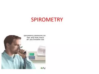
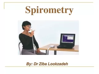
Spirometry: Airway Function Tests VC: Volume The word spirometry means “the measuring of breath.” It is the most common of the Pulmonary Function Tests (PFTs). It measures lung function, specifically the direct measurement of the amount (volume) and/or speed (flow) of air that can be inhaled and exhaled. FVC: Volume & Flow
Spirometry: Airway Function Tests • Vital Capacity (VC) • Forced Vital Capacity • Flow Volume Loop • Pre/Post Bronchodilator • Pre/Post Bronchochallenge
Spirometry: Airway Function Tests • Maximum Voluntary Ventilation (MVV) • Maximal Inspiratory (MIP) • Expiratory Pressure (MEP) • Airway Resistance (Raw) • Compliance (CL)
Indications for Spirometry • Detect the presence of lung disease Spirometry is recommended as the “Gold Standard” for diagnosis of obstructive lung disease by: • National Lung Health Education Program (NLHEP) • National Heart, Lung and Blood Institute (NHLBI) • World Health Organization (WHO)
Indications for Spirometry BOX 1-2 • Diagnose the presence or absence of lung disease • Quantify the extent of known disease on lung function • Measure the effects of occupational or environmental exposure
Indications for Spirometry BOX 1-2 • Determine beneficial or negative effects of therapy • Assess risk for surgical procedures • Evaluate disability or impairment • Epidemiologic or clinical research involving lung health or disease
SPIROMETRY • Vital Capacity The vital capacity (VC) is the volume of gas measured from a slow, complete expiration after a maximal inspiration, without a forced effort.
SPIROMETRY • Vital Capacity
SPIROMETRY • Vital Capacity • Valid VC measurements important • IC and ERV used to calculate RV and TLC • Example: • RV = FRC - ERV • TLC = IC + FRC
SPIROMETRY • VC: Criteria for Acceptability 1. End-expiratory volume varies by less than 100 ml for three preceding breaths 2. Volume plateau observed at maximal inspiration and expiration
SPIROMETRY • VC: Criteria for Acceptability 3. Three acceptable VC maneuvers should be obtained; volume within 150 ml. 4. VC should be within 150 ml of FVC value
SPIROMETRY • VC: Selection Criteria The largest single value from at least 3 acceptable maneuvers should be reported
SPIROMETRY • VC: Significance/Pathophysiology • Decreased VC • Loss of distensible lung tissue • Lung CA • Pulmonary edema • Pneumonia • Pulmonary vascular congestion • Surgical removal of lung tissue • Tissue loss • Space-occupying lesions • Changes in lung tissue
SPIROMETRY • VC: Significance/Pathophysiology • Decreased VC • Obstructive lung disease • Respiratory depression or neuromuscular disease • Pleural effusion • Pneumothorax • Hiatal hernia • Enlarged heart
SPIROMETRY • VC: Significance/Pathophysiology • Decreased VC • Limited movement of diaphragm • Pregnancy • Abdominal fluids • Tumors • Limitation of chest wall movement • Scleraderma • Kyphoscoliosis • Pain
Predicted Values • Laboratory Normal Ranges • Laboratory tests performed on a large number of normal population will show a range of results
Predicted Values • Laboratory Normal Ranges
Predicted Values • Laboratory Normal Ranges • Most clinical laboratories consider two standard deviations from the mean as the normal range since it includes 95% of the normal population.
PFT Reports • When performing PFT’s three values are reported: • Actual – what the patient performed • Predicted – what the patient should have performed based on Age, Height, Sex, Weight, and Ethnicity • % Predicted – a comparison of the actual value to the predicted value
PFT Reports • Example ActualPredicted%Predicted VC 4.0 5.0 80%
SPIROMETRY • VC: Significance/Pathophysiology • If the VC is less than 80% of predicted: FVC can reveal if caused by obstruction
SPIROMETRY • VC: Significance/Pathophysiology • If the VC is less than 80% of predicted: Lung volume testing can reveal if caused by restriction
SPIROMETRY • Forced Vital Capacity (FVC) The maximum volume of gas that can be expired when the patient exhales as forcefully and rapidly as possible after maximal inspiration (sitting or standing)
SPIROMETRY • FVC(should be within 150 ml of VC)
SPIROMETRY • FVC: Criteria for Acceptability • Maximal effort; no cough or glottic closure during the first second; no leaks or obstruction of the mouthpiece. • Good start-of-test; back extrapolated volume <5% of FVC or 150 ml, whichever is greater
SPIROMETRY • FVC: Criteria for Acceptability • Tracing shows 6 seconds of exhalation or an obvious plateau (<0.025L for ≥1s); no early termination or cutoff; or subject cannot or should not continue to exhale
SPIROMETRY • FVC: Criteria for Acceptability • Three acceptable spirograms obtained; two largest FVC values within 150 ml; two largest FEV1 values within 150 ml
SPIROMETRY • FVC: Selection Criteria The largest FVC and largest FEV1 (BTPS) should be reported, even if they do not come from the same curve
SPIROMETRY • FVC: When to call it quits !!! If reproducible values cannot be obtained after eight attempts, testing may be discontinued
SPIROMETRY • FVC: Significance and Pathophysiology • FVC equals VC in healthy individuals • FVC is often lower in patients with obstructive disease
SPIROMETRY • FVC: Significance and Pathophysiology • FVC can be reduced by: • Mucus plugging • Bronchiolar narrowing • Chronic or acute asthma • Bronchiectasis • Cystic fibrosis • Trachea or mainstem bronchi obstruction
SPIROMETRY • FVC: Significance and Pathophysiology • Healthy adults can exhale their FVC within 4 – 6 seconds • Patients with severe obstruction (e.g., emphysema) may require 20 seconds, however, exhalation times >15 seconds will rarely change clinical decisions
SPIROMETRY • FVC: Significance and Pathophysiology • FVC is also decreased in restrictive lung disease • Pulmonary fibrosis • dusts/toxins/drugs/radiation • Congestion of pulmonary blood flow • pneumonia/pulmonary hypertension/PE • Space occupying lesions • tumors/pleural effusion
SPIROMETRY • FVC: Significance and Pathophysiology • FVC is also decreased in restrictive lung disease • Neuromuscular disorders, e.g, • myasthenia gravis, Guillain-Barre • Chest deformities, e.g, • scoliosis/kyphoscoliosis • Obesity or pregnancy
SPIROMETRY • Forced Expiratory Volume (FEV1) The volume expired over the first second of an FVC maneuver
SPIROMETRY • Forced Expiratory Volume (FEV1) • FEV1 is the most widely used spirometric parameter, particularly for assessment of airway obstruction
SPIROMETRY • Forced Expiratory Volume (FEV1) • FEV1 is used in conjunction with FVC for: • Simple screening • Response to bronchodilator therapy • Response to bronchoprovocation • Detection of exercise-induced bronchospasm
SPIROMETRY • Forced Expiratory Volume (FEV1) • May be reduced in obstructive or restrictive patterns, or poor patient effort
SPIROMETRY • Forced Expiratory Volume (FEV1) • In obstructive disease, FEV1 may be decreased because of: • Airway narrowing during forced expiration • emphysema • Mucus secretions • Bronchospasm • Inflammation (asthma/bronchitis) • Large airway obstruction • tumors/foreign bodies
SPIROMETRY • Forced Expiratory Volume (FEV1) • The ability to work or function in daily life is related to the FEV1 and FVC • Patients with markedly reduced FEV1 values are more likely to die from COPD or lung cancer
SPIROMETRY • Forced Expiratory Volume (FEV1) • FEV1 may be reduced in restrictive lung processes • Fibrosis • Space-occupying lesions • Neuromuscular diseases • Obesity • Chest wall deformity
SPIROMETRY • Forced Expiratory Volume Ratio (FEVT%) • FEVT% = FEVT/FVC x 100 • Useful in distinguishing between obstructive and restrictive causes of reduced FEV1 values
SPIROMETRY • Forced Expiratory Volume Ratio (FEVT%) • Normal FEVT% Ratios for Health Adults • FEV 0.5% = 50%-60% • FEV 1% = 75%-85% • FEV 2% = 90%-95% • FEV 3% = 95%-98% • FEV 6% = 98%-100% • Patients with obstructive disease have reduced FEVT% for each interval
SPIROMETRY • Forced Expiratory Volume Ratio (FEVT%) • A decrease FEV1/FVC ratio is the “hallmark” of obstructive disease • FEV1/FVC <75%
SPIROMETRY • Forced Expiratory Volume Ratio (FEVT%) • Patients with restrictive disease often have normal or increased FEVT% values • FEV1 and FVC are usually reduced in equal proportions • The presence of a restrictive disorder may by suggested by a reduced FVC and a normal or increased FEV1/FVC ration
SPIROMETRY • Forced Expiratory Flow 25% - 75% (maximum mid-expiratory flow) • FEF 25%-75% is measured from a segment of the FVC that includes flow from medium and small airways • Normal values: 4 – 5 L/sec
SPIROMETRY • Forced Expiratory Flow 25% - 75% In the presence of a borderline value for FEV1/FVC, a low FEF 25%-75% may help confirm airway obstruction