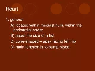
Heart
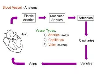
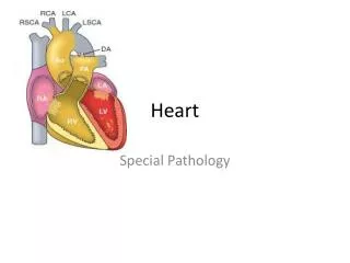
Heart . Pericardium. 1. visceral = inseparatable with epicardium 2. parietal = outer layer of sac 3. pericardial cavity . Major vessels . all covered by visceral pericardium pulmonary trunk - exits right ventricle from anterior surface


Heart
E N D
Presentation Transcript
Pericardium • 1. visceral = inseparatable with epicardium2. parietal = outer layer of sac3. pericardial cavity
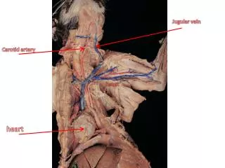
Major vessels • all covered by visceral pericardium • pulmonary trunk - exits right ventricle from anterior surface • aorta - exits left ventricle just posterior to pulmonary trunk • superior vena cava - enters right atrium on right, posterior to aorta • inferior vena cava - enters right atrium, inferior to superior vana cava • pulmonary veins - (4) upper & lower on right & left - enter left atrium, posterior side of heart
Surface Structure of Chambers • Right Ventricle - forms majority of anterior (sternocostal) surface • Right Atrium - forms right lateral surface • auricle - pointed tissue appendage on surface of both atria • atrioventricular (coronary) sulcus - groove separates atrium & ventricle - holds coronary artery, sinus
Surface Structure of Chambers • Left Ventricle - forms left side of anterior surface, most of posterior • a. anterior, posterior interventricular sulcus - separate right, left ventricles - hold coronary artery and vein • b. apex - pointed inferior end of heart, • Left Atrium - most superior • base of heart
Inner structure of Chambers • Wall layers - all chambers • endocardium - inner layer lining chambers - continuous with vessel endotheliu • myocardium - bulk of muscle walls • epicardium – inseparable with visceral pericardium
Right atrium • crista terminalis - ridge of muscle connects superior and inferior vana cava (= sulcus terminalis on surface) • Pectinate muscle - small muscle ridges inside anterior wall of right atrium • sinus venarum - smooth surfaced area where veins enter right atrium entrances of superior and inferior vana cava, coronary sinus • Right atriventricular orifice, tricuspid valve • interatrial septum, fossa ovalis - remnant of foramen ovale (in fetus) *check if patent
1. Sinus venarum 2. Terminal crest 3. Fossa ovalis 4. Rim or limbus of the fossa ovalis 5. Musculi pectinati 6. Valve of the inferior vena cava
Right ventricle • anterior wall = sternocostal surface • medial wall = Interventricular septum • inferior wall = diaphragmatic surface • conus arteriosus /infundibulum - superior part of right ventricle, narrows toward pulmonary trunk • opening to pulmonary trunk, pulmonary valve - behind 3d left costal cartilage • Right atrioventricular orifice, tricuspid valve - attached at peripheral edge to anulus fibrosus (ring) • trabeculae carneae - ridges of muscle on inner surface - except in conus arteriosus • chordae tendineae - fibrous tissue, attach ends of valve cusps to papillary muscles
Left atrium • 4 openings of pulmonary veins • Left atrioventricular orifice, mitral (bicuspid) valve • Pectinate muscle, interatrial septum, fossa ovalis
Left ventricle • aortic valve: three cusp, 3rd intercostal space • ascending aorta - (becomes aortic arch behind sternal angle) • R, L coronary arteries - exit ascending aorta just distal to valve cusps • Left atrioventricular orifice, bicuspid valve, anulus fibrosus, chordae tendineae, papillary muscles • Trabeculae carneae - except in aortic vestibule ( smooth region, fibrous and leads to ascending aorta)
Coronary vessels • Right coronary artery - in coronary sulcus; travels to the right & inferiorly • supplies right atrium, right ventricle, posterior part of interventricular septum, SA and AV node, AV bundle; also part of left atrium and left ventricle • Right marginal artery – runs towards the apex of the heart • posterior interventricular artery - sits in posterior interventricular sulcus, supplies both ventricles (anastomoses with anterior interventricular artery) • most likely (85%) an atrioventricular (AV) nodal artery
Coronary vessels • Left coronary artery - curves left in coronary sulcus, divides quickly • supplies left atrium, left ventricle, anterior IV septum, part of right atrium • anterior interventricular artery - in anterior interventricular sulcus, to the apex of the heart, supplies both ventricles and interventricular septum • circumflex artery. - continues left in coronary sulcus around to posterior of heart, supplies left atrium, base of left ventricle • Left marginal artery, branch of circumflex
Coronary vessels • The branches of coronary artery are end branches. They supply regions of cardiac muscle without overlap of other large branches. Anastomoses are generally not large enough for survival. • Large branch occluded distal region ischemic necrosis MI • Risk factor: Coronary atherosclerosis
Coronary vessels • Angina pectoris: • Exertion, stress, exercise after meal • Most common sites for coronary occlusion: • The anterior interventricular branch of the left coronary artery • The right coronary artery • The circumflex artery of left coronary artery • Treatment: Bypass operation
Veins • Great cardiac vein - in anterior interventricular sulcus - around left side in coronary sulcus, empties into coronary sinus (along with anterior interventricular artery) • Middle cardiac vein - in posterior interventricular sulcus - also empties into coronary sinus (along with posterior interventricular artery) • Small cardiac vein - around right side in coronary (with marginal branch of the right coronary artery)
Veins • coronary sinus - largest coronary vein; in posterior coronary groove, drains into right atrium