Download
1 / 70
820 likes | 1.57k Vues
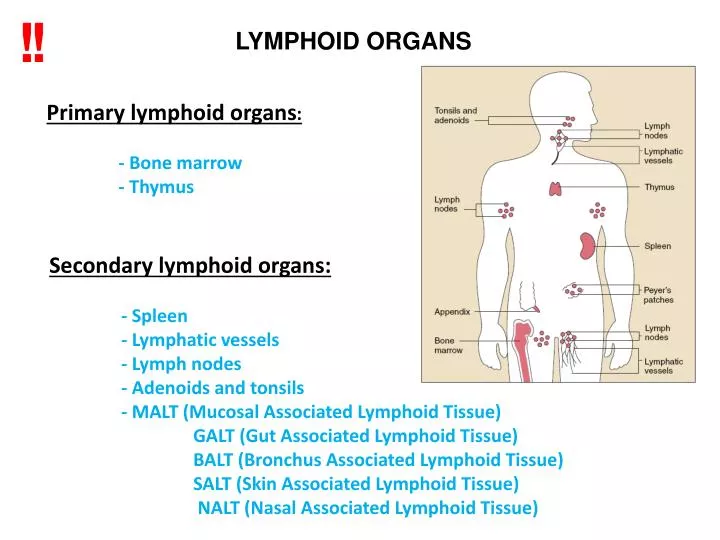
!. !. LYMPHOID ORGANS. Prim a r y lymphoid organs : - B one marrow - T hymus. Secondary lymphoid organs: - S pleen - Lymphatic vessels - L ymph node s - Adenoids and t onsils - MALT (Mucosal Associated Lymphoid Tissue) GALT (Gut Associated Lymphoid Tissue)

E N D
! ! LYMPHOID ORGANS Primary lymphoid organs: - Bone marrow - Thymus Secondary lymphoid organs: - Spleen - Lymphatic vessels - Lymph nodes - Adenoids and tonsils - MALT (Mucosal AssociatedLymphoid Tissue) GALT (Gut Associated Lymphoid Tissue) BALT (Bronchus Associated Lymphoid Tissue) SALT (Skin Associated Lymphoid Tissue) NALT (Nasal Associated Lymphoid Tissue)
! ! THE TWO ARMS OF THE IMMUNE SYSTEM Monocytes, Macrophages, Dendritic cells, Granulocytes, NK cells and Complement components Monocytes, Macrophages, Dendritic cells, Granulocytes, NK cells and Complement components B and T cells
! ! Professional phagocytic cells macrophages neutrophyl granulocytes dendrtitic cells the phagocytosed cells or molecules may modify the functions of the cell phagocytosis followed by enzymatic degradation Professional antigen presenting cells macrophages B lymphocytes dendrtitic cells they express MHCII molecules the protein degradation products (peptides) can be presented to T lymphocytes by MHC molecules ! !
Cells of innate immune system: • Macrophages: • Macrophages are constitutively present in tissues and recognize microbes that enter these tissues and respond rapidly to these microbes. Initiate the immune response • These cells are phagocytes (eliminate the pathogens) • Activate the innate immune response(bysecreted proteins, called cytokines) • Activate the adaptive immune system. Macrophages serve as APCs that display antigens to and activate T lymphocytes • Dendritic cells • are constitutively present in tissues and recognizerapidly microbes that enter these tissues. Initiate the immune response. • They have phagocytic capabilities • migrate to lymph nodes, and display microbial antigens to T lymphocytes,professional antigen presentimg cells (APC) • Neutrophil granulocytes are phagocytes, the main function toeliminate the pathogens • Appear only in the circulation under normal condition Main actors In inflammatory processes ! !
Phagocyte activation How do immunocytes communicate: Soluble mediators Infection CYTOKINES & CHEMOKINES Soluble proteinsproduced by cells. They have strong effect on the function of other cells. Bit similar to hormones.
CTL Cell killing Target cell T Antigen presentation Y T B Antibody production Activation of accessory cells Dendritic cell macrophage How do immunocytes communicate: Cell-cell interaction Cell-cell communication takes place commonly in all the phases of the immune response
THE MOST IMPORTANT FEATURES OF CYTOKINES ! • The most important mediators of indirect cell communication in the immune system („hormones” of the immune system). • Act in low concentrations. • Cytokines can affect in an autocrine way, in a paracrine way, • or in an endocrine way • pleiotropic effect. • Cytokines can act by synergistic or antagonistic ways to each other. • A given cell may by affected by many cytokines resulting in the • same effect redundant effect. • The responsiveness of the given cell is based on the expression of cytokine-specific receptors.
Cytokines can be devided into sub-groups by origin and functional properties. Functional groups: Inflammatory cytokines Direct the development and maturation of immune cells Direct activation and differentiation of immune cells
Categories of cytokines hormons cytokines interleukins chemokine interferons
MOLECULES OF THE IMMUNE SYSTEM • Most important receptors of the imune system • receptors (BCR, TCR,MHCI, MHCII, PRR, etc.) • Soluble molecules: • cytokines • antibodies • complement components
Receptors responsible for the recogniton of pathogens in the immune system
Physical and chemical barriers Stomach Respiratory tract • pH of 3-4 • Pepsin • Cilliary movement • Coughing, sneezing Skin Impaired cilia movement (CF)! • Tight junctions • Keratin layer • Antibacterial peptides; Defensins • pH of 5.5 • Fatty acids Burns and susceptibility to infections! Eye • Tear film (Oils, lactoferin, mucin and lyzosyme) Vagina • pH of 3.8-4.5 • Lactobacillus Lactic acid
INNATE IMMUNITY Pathogen recognition PRRs (TLRs, C type lectins, Mannose and Glucan binding lectins, NLRs and RIG-I helicases) Phagocytosis, effector functions Communication/ Antigen presentation Intracellular – on surface MHC I complex proteins Extracellular – on surface MHC II complex proteins
! ! Innate immunity as a first line of defence Innate immune cells recognize frequently found structures of pathogens, these are not found in human cells! Examples: duple strain RNA bacterial cell wall components bacterial flagellin…. Recognition is inevitable
! ! Danger signal! The innate immune system also recognizes molecules that are released from damaged or necrotic cells. Such molecules are called damage-associated molecular patterns (DAMPs).
! ! Innate immunity as a first line of defence Innate immune cells recognize frequently found structures of pathogens, these are not found in human cells! Examples: duple strain RNA bacterial cell wall components bacterial flagellin…. Recognition is inevitable
PAMPs- Pathogen associated molecular patters Structures on pathogens recognized by the innate cells
PRR types TOLL RIG like receptors NOD Scavanger receptors C type lectin receptors Mannose recognizing receptors
Virus Bacteria ssRNS dsRNA CpG DNA Gram- Flagellin Peptidoglycane LPS Gram+ TLR9 TLR7 TLR8 TLR3 TLR2 TLR6 TLR5 TLR4 Interferon producing cell PC/DC IFN Macrophage/Dendritic cell TLRs RECOGNIZE VARIOUS MICROBIAL STRUCTURES
TLR receptors: • Intracellular and cell surface sensors. • Viral RNA, non-methylated DNA characteristic of bacteria, bacterial flagella, bacterial • surface components (lipoproteins, peptidoglicans) and fungi structures. • Partial overlapping recognition between NOD and RIG like receptors.
NOD like receptors • NOD-like receptors: • Intracellular receptors. • Recognizing intracellular pathogen and danger signals. • Partial overlapping recognition with TLRs.
RIG receptors: • Intracellular sensors. • Recognizing viral RNA, inducing an anti-viral response. • Partial overlapping recognition with TLRs.
Eukaryotes Mannose Bacterium Mannóz Mannose receptors Prokaryotes Glucosamine Galactose Mannose Siallic acid Macrophage / Dendritic cell
Specificity of innate immunity ! direct connetion between innate cells and pathogen ( ) Few receptors (20-30) are responsible for the recognition of all the pathogens
! OPSONIZATION ! Opsonization facilitate and accelerate the recognition of the pathogen by phaogocytes, opsonins form a bridge between pathogen and a phagocyte connecting them. Main opsonins: antibodies Complement fragments Acute-phase proteins
Pathogen recognition by innate immune system • Directly via PRR • Indirectly via opsonization
INNATE IMMUNITY Pathogen recognition PRRs (TLRs, C type lectins, Mannose and Glucan binding lectins, NLRs and RIG-I helicases) Effector functions, elimination of pathogens Communication/ Antigen presentation Intracellular – on surface MHC I complex proteins Extracellular – on surface MHC II complex proteins
! ! • INNATE IMMUNITY II • Effector functions, elimination of pathogens • Phagocytosis • Killing with soluble mediators • Complement system • NK cell activation
Degradation PRR ACTIVATION Bacterium Intracellular killing Phagocyte Uptake Antigen presentation T cell ACQUIRED IMMUNITY PHAGOCYTOSIS 0.5 - 1 hours The amount of internalized particles is limited
THE PHAGOCYTIC SYSTEM MACROPHAGES DENDRITIC CELLS NEUTROPHILS
Phagocytic cells • Macrophages • Dendritic cells • Neutrophil granulocytes • (No presentation on MHC II) • Professional • antigen presenting cells • Macrophages • Dendritic cells • B lymphocytes • (no killing action, only Ag presentation)
Extracellular pathogen phagocytosis and killing
Extracellular pathogen phagocytosis and killing
2. Soluble mediators reeased from macrophages, granulocytes are responsible for kiliing of extracellular pathogens ROS reactive oxigen species NO nitric oxide Destructive enzymes, antimicrobial substances
Intracellular bacterial evasion of killing in phagocytes Macrophage effector capacity Defensins Phagosome acidification Phagosome–lysosome fusion Lysosomal enzymes Intraphagolysosomal killing ROI RNI Iron starvation Tryptophan starvation
3. COMPLEMENT ACTIVATION COMPLEMENT Lysis of bacteria Complement-proteins Inflammation Chemotaxis Bacterium Lectin pathway Alternative pathway Complement-dependent phagocytosis Antigen + Antibody ACQUIRED IMMUNITY Few minutes – 1 hour Enzymes get fragmented, complement activity can be exhausted
NK cells • Major differences between NK cells and B/T lymphocytes: • Contain large cytoplasmic granules. • Responds fast, circulate in a partly activated state. • Do not express surface receptors produced by rearranged genes. • Have a range of cell-surface receptors that deliver activating or inhibitory signals • Have two main types of receptors: Ig-like Rs and the Lectin-like Rs (inhibitory and activating) that recognize altered cell surface proteins as a result of a virus infection. • Overall balance of inhibitory or activating signals decides if the NK cell killing action will take place. • Individual NK cells express different combinations of receptors- heterogeneity repertoire of responses to pathogens.
! Killing of the cells infected with intracellular pathogens Target MHC+ Target MHC- KIR KIR KAR KAR NK NK KIR – Killer Inhibitory Receptor association to MHC I KAR – Killer Activatory Receptor The activity of NK cells is enhanced by activatory receptors Inhibitory receptors block NK cell activity. Self cells are protected by inhibitory receptors. Infection or tumors may increase the amount of activation and/or decrease the efficacy of inhibition Inhibition of lysis lysis