Download
1 / 2
20 likes | 83 Vues
Supplementary Figure 1. A. B. #1 #2 #3. Group 1. #4 #5 #6. CD20. Group 2. C. #7 #8 #9. Group 3. MHC I. Her-2/ neu.

E N D
Supplementary Figure 1 A B #1 #2 #3 Group 1 #4 #5 #6 CD20 Group 2 C #7 #8 #9 Group 3 MHC I Her-2/neu
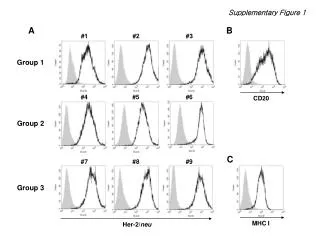
Supplementary Figure 1. Confirmation of Her-2/neu expression on autologous B lymphoblastoid cells (BLCL) transduced with retroviral vector expressing Her-2/neu. A. Autologous BLCL and BLCL-HM were stained with PE-conjugated anti-Her-2/neu antibody. Binding of anti-Her-2/neu Ab was analyzed by flow cytometry. BLCL is shown as a filled area, and BLCL-HM is shown as a solid line. B. BLCL #1 was stained with FITC-conjugated anti-human CD20 antibody. CD20 expression is shown as a solid line. Binding of the control antibody is shown as a filled area. C. BLCL #1 was stained with PE-conjugated anti-human MHC I antibody. The level of MHC I is shown as a solid line. Binding of the control antibody is shown as a filled area. Data are representative of at least three separate experiments.