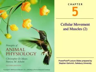
Understanding Cellular Movement: The Role of Cytoskeleton and Motor Proteins
This article explores the intricate dynamics of cellular movement, emphasizing the importance of the cytoskeleton and motor proteins. It details the types of cellular organisms, including sponges, cnidarians, and various invertebrates, and outlines the structural components of the cytoskeleton—microtubules and microfilaments—along with their assembly and regulation processes. Key mechanisms such as intracellular transport, motility, and changes in cell shape are discussed, illustrating how different motor proteins like kinesin and dynein facilitate movement and maintain cell integrity in various organisms.

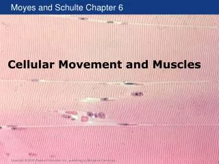
Understanding Cellular Movement: The Role of Cytoskeleton and Motor Proteins
E N D
Presentation Transcript
Cnidarians Sponges (phylum Porifera) Ctenophores Hydra Medusa Sea gooseberries Sea walnut
Nematodes Flatworms Annelids Annelids – Internal structure
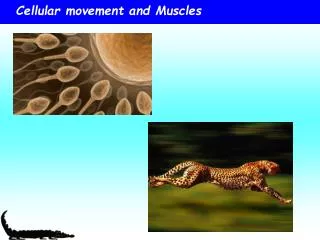
Cytoskeleton and Motor Proteins • All physiological processes depend on movement • Intracellular transport • Changes in cell shape • Cell motility • Animal locomotion
Cytoskeleton and Motor Proteins • All movement is due to the same cellular “machinery” • Cytoskeleton • Protein-based intracellular network • Motor proteins • Enzymes that use energy from ATP to move
Use of Cytoskeleton for Movement • Cytoskeleton elements • Microtubules • Microfilaments
Reorganizing the cytoskeletal network A macrophage of a mouse stretching its arms to engulf two particles, possibly pathogens
Cytoskeleton and Motor Protein Diversity Structural and functional diversity Alteration of function Multiple isoforms Various ways of organizing
Microtubules • Are tubelike polymers of the protein tubulin • Similar protein in diverse animal groups • Multiple isoforms • Are anchored at both ends • Microtubule-organization center (MTOC) (–) near the nucleus • Attached to integral proteins (+) in the plasma membrane
Function of Microtubules • Motor proteins can transport subcellular components along microtubules • Motor proteins kinesin and dynein • For example, rapid change in skin color
Microtubules: Composition and Formation • Microtubules are polymers of the protein tubulin • Tubulin is a dimer of a-tubulin and b-tubulin • Tubulin forms spontaneously • For example, does not require an enzyme • Polarity • The two ends of the microtubule are different • Minus (–) end • Plus (+) end
Microtubule Assembly • Activation of tubulin monomers by GTP • Monomers join to form tubulin dimer • Dimers form a single-stranded protofilament • Many protofilaments form a sheet • Sheet rolls up to form a tubule • Dimers can be added or removed from the ends of the tubule • Asymmetrical growth • Growth is faster at + end • Cell regulates rates of growth and shrinkage
Microtubule Growth and Shrinkage Dynamic instability MAPs Temperature Local [tubulin] Growth / Shrinkage Chemicals (Taxol, Colchicine) STOPs Katanin GTP hydrolysis on b-tubulin
Microtubule Dynamics Figure 5.5
Regulation by MAPs Figure 5.6
Movement Along Microtubules • Motor proteins move along microtubules • Direction is determined by polarity and the type of motor protein • Kinesin move in (+) direction • Dynein moves in (–) direction • Movement is fueled by hydrolysis of ATP • Rate of movement is determined by the ATPase domain of motor protein and regulatory proteins • Dynein is larger than kinesin and moves five times faster
Vesicle Traffic in a Neuron Figure 5.7
Cilia and Flagella • Cilia • numerous, • wavelike motion. • Flagella • single or in pairs, • whiplike movement.
Microtubules and Physiology Table 5.1
Microfilaments • Polymers composed of the protein actin • Found in all eukaryotic cells • Often use the motor protein myosin • Movement arises from • Actin polymerization • Sliding filaments using myosin • More common than movement by polymerization
Microfilament Structure and Growth • G-actin monomers polymerize to form a polymer called F-actin • Spontaneous growth • 6–10 times faster at + end • Treadmilling • Assembly and disassembly occur simultaneously and overall length is constant • Capping proteins • Increase length by stabilizing – end and slowing disassembly
Microfilament (Actin) Arrangement • Arrangement of microfilaments in the cell • Tangled neworks • Microfilaments linked by filamin protein • Bundles • Cross-linked by fascin protein • Networks and bundles of microfilaments are attached to cell membrane by dystrophin protein • Maintain cell shape • Can be used for movement
Microfilament (Actin) Arrangement Figure 5.10
Movement by Actin Polymerization • Two types of amoeboid movement • Filapodia are rodlike extensions of cell membrane • Neural connections • Microvilli of digestive epithelia • Lamellapodia are sheetlike extensions of cell membrane • Leukocytes • Macrophages
Actin Polymerization and Fertilization Figure 5.11
Myosin • Most actin-based movements involve the motor protein myosin • Sliding filament model • 17 classes of myosin (I–XVII) • Multiple isoforms in each class • All isoforms have a similar structure • Head (ATPase activity) • Tail (can bind to subcellular components) • Neck (regulation of ATPase)
Sliding Filament Model Sliding Filament Model Chemical reaction Structural change Myosin binds to actin (cross-bridge) Myosin bends (power stroke) • Myosin is an ATPase • Converts energy from ATP to mechanical energy • Need ATP to release and reattach to actin • Absence of ATP causes rigor mortis • Myosin cannot release actin
Sliding Filament Model - Cross-bridge cycle Extension Cross-bridgeformation Power stroke Release Figure 5.13
Actino-Myosin Activity • Two factors affect movement • Unitary displacement • Distance myosin steps during each cross-bridge cycle • Depends on • Myosin neck length • Location of binding sites on actin • Helical structure of actin • Duty cycle • Cross-bridge time/cross-bridge cycle time • Typically ~0.5 • Use of multiple myosin dimers to maintain contact
Myosin Activity Figure 5.14
Actin and Myosin Function Table 5.2