Download

1 / 20
240 likes | 1.2k Vues
LIQUIDE AMNIOTIQUE ET PLACENTA. COURS ESF 2006. LE PLACENTA. PLACENTA. L’Echographie permet une exploration placentaire inaccessible à la clinique Elle a apporté un bénéfice considérable dans des décisions obstétricales. 1. ECHOGRAPHIE PLACENTA IMAGE NORMALE. PLACENTA.

E N D
LIQUIDE AMNIOTIQUE ET PLACENTA COURS ESF 2006
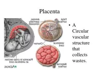
PLACENTA • L’Echographie permet une exploration placentaire inaccessible à la clinique • Elle a apporté un bénéfice considérable dans des décisions obstétricales.
1. ECHOGRAPHIE PLACENTA IMAGE NORMALE
PLACENTA 1° TRIMESTRE
ASPECTS ECHOGRAPHIQUE DU PLACENTA DEBUT GSS • Placenta = Throphoblaste • Visualisation sous forme de couronne throboblastique échogène uniforme • S’ épaissit à partir de 10 sem pour se localiser dans la zone ou se trouvera le placenta 14 sem
PLACENTA 2° TRIMESTRE
ASPESCT ECHO PLACENTA • Echogène gris moyen • Forme limitée • côté amniotique par une zone échogène • plaque choriale • côté utérin limité par fin liseré • plaque basale
PLAQUE CHORIALE CORDON plaque choriale plaque basale
PLACENTA • A partir de 20 sem apparaissent des espaces anéchogènes sinus veineux ( signal doppler + ) • a différencier d’un décollement ( signal doppler -)
LOCALISATION PLACENTAIRE 2 ° TRIM • Placenta sera localisé : • * Placenta antérieur • * Placenta postérieur
PLACENTA PRAEVIA LOCALISATION PLACENTAIRE 2 ° TRIM • Placenta sera localisé : sonde plan de coupe longitudinale • * Placenta antérieur : haut de l’image • * Placenta postérieur : bas de l’image
REPERAGE PLACENTA / O.IDU COL • Primordial à l’échographie du 2° trimestre
LOCALISATION PLACENTAIRE • Position placenta / mur de la vessie • Atteint le mur • N’atteint pas le mur de la vessie
LOCALISATION PLACENTAIRE • BAS INSERE • NON BAS INSERE
PLACENTA • 3° TRIMESTRE