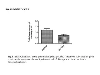
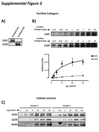
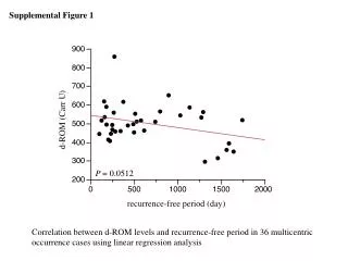
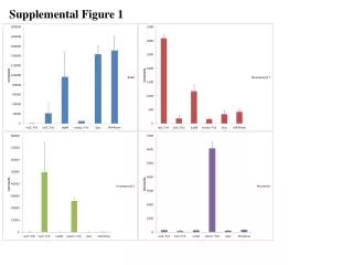
Download
1 / 6

E N D
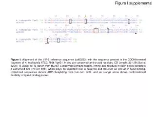
Figure I supplemental Figure I: Alignment of the VIP-2 reference sequence (cd00233) with the sequence present in the COOH-terminal fragment of A. hydrophila ATCC 7966 VgrG1. In red are conserved amino-acid residues. CD Length: 201; Bit Score: 62.27; E-value: 5e-10 (taken from BLAST-Conserved Domains report). Amino acid residues in open boxes constitute a conserved Ser-Thr-Ser motif, which plays an important role in catalysis and structure as well as in NAD binding. Underlined sequences denote ADP-ribosylating toxin turn-turn motif, and an orange arrow shows conformational flexibility of ligand binding pocket.
Figure II Supplemental A. Phage_GPD DUF586 VIP-2 VgrG COOH-terminal NH2-terminal 1aa 701aa 927aa B. Phage_GPD DUF586 RtxA VgrG COOH-terminal NH2-terminal 1163aa 701aa 1aa gp27-like (1 to554aa) gp5-like (554 to 650 aa) Figure II: Schematic representation of the conserved domains present in VgrG1. A.A. hydrophila ATCC 7966 (gi|117619461) and B.V. cholerae N16961 (gi|15641427). The gp27- and gp5-like motifs are represented by red and blue lines, respectively (modified from Pukatzki, et al 2007) (38). The encoding fragments representing the NH2-terminal (gray), COOH-terminal (cyan) and the full-length (gray and cyan) of VgrG1 from A. hydrophila ATCC 7966 were cloned into a pET-30a vector to produce recombinant proteins, and into pBI-EGFP vector for expression in the HeLa Tet-off cells. The positions of VIP-2 and RtxA domains in VgrG1 of A. hydrophila and V. cholerae are also shown.
Figure III Supplemental act Control act/ vasH pBR322-empty act/ vasH pBR322-vasH A.
Figure III Supplemental Pre Post Δact ΔvasH Δact ΔvasH Δact Δact VgrG 2/3 (~77 kDa) Hcp (~20 kDa) 1 2 3 4 B. Figure III: A. Induction of HeLa cell-rounded phenotype in co-cultures with different strains of A. hydrophila SSU. HeLa cells were co-cultured in direct bacterial-host cell contact for 90 min with A. hydrophila SSU Δact mutant (Top right), Δact/ΔvasH pBR322-empty (with vector alone) (Bottom left), and Δact/ΔvasH pBR322-vasH in which the Δact/ΔvasH mutant of A. hydrophila SSU was complemented with the vasH gene using the pBR322 vector in trans(Bottom right). Normal morphology of HeLa cells is shown in Top left panel (control). Magnification 40X. B. Detection of Hcp2 and VgrG 2/3 in tissue culture supernatants after co-culturing of HeLa cells with different strains of A. hydrophila SSU. Pre (lanes 1-2): filtered tissue culture supernatants after 90 min of co-culture of HeLa cells with bacteria and before they were used as pre-conditioned media on fresh HeLa cells. Post (lanes 3-4): same conditioned tissue culture supernatants after 120 min on HeLa cell cultures.
VgrG1-NH2::Bla VgrG1-NH2::Bla VgrG1-Full::Bla VgrG1-Full::Bla Empty Empty ~131 kDa Pellets ~106 kDa ~131 kDa ~106 kDa Supernatants 1 2 3 4 5 6 act act/vasH Figure IV Supplemental Figure IV: Western blot analysis of A. hydrophila SSU act(lanes 1 to 3) and act/vasH(lanes 4 to 6) mutant bacterial pellets (top panel) and supernatants (bottom panel) using antibodies to Bla. The production of full-length VgrG1 (VgrG1-Full::Bla) (lane 2) and VgrG1-NH2::Bla (lane 3) was detected in both bacterial pellet and supernatant of A. hydrophila SSU act parental strain. In the act/vasH mutant strain, fusion proteins were detected in the pellets (lanes 5 and 6) but not in the supernatants. Bacterial strains transformed with the empty vector were used as a control (lanes 1 and 4).
Figure V Supplemental pBI-EGFP empty pBI-EGFP vgrG2 Figure V: Morphological changes of HeLa Tet-off cells induced by theexpression of the vgrG2 gene of A. hydrophila SSU. Cells were stained for actin-cytoskeleton by using Alexa fluor 568-phalloidin (red), and expression of the gene encoding EGFP was detected in HeLa cells successfully transfected with the pBI-EGFP vector alone (Left) or containing the vgrG2 gene (Right). Magnification 40X.