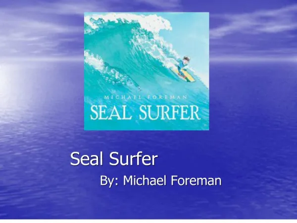
Using 3D-SURFER
Using 3D-SURFER. Before you start. 3D-Surfer can be accessed at http://dragon.bio.purdue.edu/3d-surfer For visualization of results, 3D-Surfer requires a Java enabled browser. The Java plugin can be downloaded from http://java.sun.com/products/plugin/downloads/

Using 3D-SURFER
E N D
Presentation Transcript
Before you start • 3D-Surfer can be accessed at http://dragon.bio.purdue.edu/3d-surfer • For visualization of results, 3D-Surfer requires a Java enabled browser. The Java plugin can be downloaded from http://java.sun.com/products/plugin/downloads/ • The Jmol package is used for visualizing protein structures. Jmol tutorials can be accessed from http://jmol.sourceforge.net/docs/
About 3D-Surfer • 3D-SURFER is a web based tool for protein surface comparison and analysis • Rapid comparison of protein structures is enabled by the use of 3D Zernike moment invariants (Novotni and Klein, 2004) • The server is easy to use. The only input is a PDB ID followed by a chain (if any). • Read on to see what 3D-Surfer can do
3D-Surfer Functionality • Search and retrieve PDB structures that have similar surface shapes based on the Euclidean distance between the descriptor vectors representing the proteins • Results displayed include • CATH codes that reflect the structural topology • Alignments based on CE can also be viewed at the click of a button • View local geometric features of interest using VISGRID • Zernike Invariants of each protein (text and graph)
Select a Protein chain Enter the protein PDB code followed by the chain ID (if any). Format “####-#”
Select a Protein chain A pop up menu assists in the selection of the protein
Select a Protein chain Optionally, change the filtering settings. CATH filtering, hits with equal CATH codes, while residue length filtering only displays matches that are similar in length to the query structure
Select a Protein chain Click Submit Click on “Submit” to view the results of the query
File Upload Access the “Upload” page Select the PDB file to be processed by uploading a structure using the “Browse” button. Add the chain ID (if any). Click on “Submit” to view the results.
The Results Page An overview of the Results page
Listing Results Results o f the query may saved separately by clicking on the “List Results” button. The user may also select the number of entries (default 25) to be listed.
Results Moving the mouse over the images produces the animated surface displaying different views. Click on the image to access query results for the PDB chain Moving the mouse over the text highlights the clickable link to the PDB website. Euclidean distance between the Zernike moments of the query protein and 1CI1-A Select the Check box to calculate the RMSD between the query protein and 1CI1 using CE. See the next slide for details
Visualizing Alignments Click on the “Rmsd” button to view the CE based structural alignment RMSD between structures as calculated by the CE program
VISGRID Click to identify Cavity regions Click to identify Protrusion regions Click to identify Flat regions Visualize the results in terms of the list of the residues forming of the three largest groups. Areas and volumes for these regions are also reported.
VISGRID Largest Cavity 2nd Largest Cavity 3rd Largest Cavity Surface area and volume of the convex hull formed by the atoms of the listed residues
Other links • Documentation - About 3D-SURFER • FAQ - Definitions of terminology and other questions on using 3D-SURFER • Please help us improve the website by sending your comments and suggestions. Access our list of contacts at http://dragon.bio.purdue.edu/3d-surfer/index.php?contact