Exploring Sliding Filament Theory: Mechanism of Muscle Contraction
630 likes | 948 Vues
Learn how the sarcomere organization and cross-bridge interactions drive muscle contraction in the Sliding Filament Theory. Discover the steps involved and the importance of ATP and calcium ions.

Exploring Sliding Filament Theory: Mechanism of Muscle Contraction
E N D
Presentation Transcript
SLIDING FILAMENT THEORY Dr. Ayisha Qureshi Assistant Professor MBBS, MPhil
Important: 3-dimensionally, thin filaments are arranged hexagonally around thick filaments and the cross-bridges project from each thick filament in all 6 directions towards the surrounding thin filaments……each thin filament is surrounded by 3 thick filaments.
What are Cross-bridges? • With an electron microscope, fine cross bridges can be seen extending from each thick filament to the thin filament. These are formed by the arm and head of the myosin molecules projecting outward from the tail, and pointing towards the thin filaments.
Questions • How does cross-bridge interaction between actin and myosin bring about muscle contraction? • How does a muscle action potential trigger this contractile process? • What is the source of the Ca2 that physically repositions troponin and tropomyosin to permit cross-bridge binding?
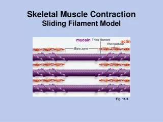
SLIDING FILAMENT THEORY Definition: When a muscle cell contracts, the thin filaments slide past the thick filaments, and the sarcomere shortens. This process comprised of several steps is called the Sliding Filament Theory. It is also called the Walk Along Theoryor the Ratchet Theory.
After the ATP has bound to the myosin head, the binding of Myosin to Actin molecule takes place:
Once the actin active sites are uncovered, the myosin binds to it:
SLIDING FILAMENT THEORY It has the following steps: • Before contraction begins, An ATP molecule binds to the myosin head of the cross-bridges. • The ATPase activity of the myosin head immediately cleaves the ATP molecule but the products (ADP+P) remains bound to the head. Now the myosin head is in a high energy state and ready to bind to the actin molecule. • When the troponin-tropomyosincomplex binds with calcium ions that come from the sarcoplasmic reticulum, it pulls the tropomyosin so that the active sites on the actin filaments for the attachment of the myosin molecule are uncovered. • Myosin head binds to the active site on the actin molecule.
SLIDING FILAMENT THEORY (cont) • The bond b/w the head of the cross bridges(myosin) & the actin filaments causes a the bridge to change shape bending 45° inwards as if it was on a hinge, stroking towards the centre of the sarcomere, like the stroking of a boat oar. This is called a POWER STROKE. • This power stroke pulls the thin filament inward only a small distance. • Once the head tilts, this allows release of ADP & phosphate ions. • At the site of release of ADP, a new ATP binds. This binding causes the detachment of the myosin head from the actin. • A new cycle of attachment-detachment-attachment begins. • Repeated cycles of cross-bridge binding, bending and detachment complete the shortening and contraction of the muscle.
ParticipantWill bind to: 1. Myosin ATP, Actin 2. Actin Myosin, Troponin 3. Tropomyosin Troponin 4. Troponin Calcium, Actin Tropomyosin 5. ATP Myosin 6. Calcium ions Troponin
Important • Through the attachment-detachment-attachment cycle, the myosin heads or cross bridges “walk” along an actin filament to pull it inward relative to the stationary thick filament. • Because of the way the myosin molecules are oriented within a thick filament, all the cross-bridges stroke towards the center of the sarcomere. • At any time during contraction, part of the cross bridges are attached to the thin filaments and are stroking, while others are returning to their original conformation in preparation for binding with another actin molecule. Thus, some cross-bridges “hold on” while others “let go”. Otherwise, the thin filaments would slip back to their resting position b/w strokes. • The detachment of the myosin head from the actin cannot take place until and unless a new ATP does not attach to the myosin head. This is important when death occurs, no more ATP is available and thus, rigor mortis occurs.
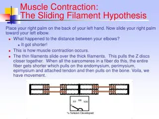
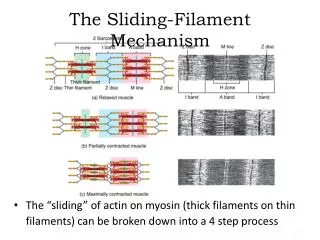
Shortening of the Muscle: The thick and thin filaments DO NOT shorten. Contraction is accomplished by the thin filaments from opposite sides of each sarcomere sliding closer together or overlapping the thick filaments further. The H-zone becomes smaller as the thin filaments approach each other. The I band becomes smaller as the thin filaments further overlap the thick filaments. The width of the A band remains unchanged as it depends on the thick filaments and the thick filaments do not change length.
½ I ½ I A When muscle contracts, the sarcomere shortens. The I band and H Zone also shorten. But the length of the A band remains the same.
Mechanism of muscle contraction: • Width of sarcomere decreases from 2.2 to 2 or less • Length of thick and thin filament remain the same • Power stroke pull the Z discs towards the center of the sarcomere • Results in app. Of the Z discs • Cyclic attachment-detachment-attachment of myosin head to actin, till there is complete overlap of thick & thin filaments • Decrease in I-band and H-band, A-band stays the same
NEUROMUSCULAR JUNCTION A NEUROMUSCULAR JUNCTION is an area of contact between a muscle fibre and a neuron. Fig. An electron micrographic sketch of the junction between a single axon terminal and the muscle fiber membrane.
MOTOR END-PLATE Definition: It is the specialized portion of a muscle fibre immediately under a terminal nerve fibre. The nerve fibre invaginates a muscle fibre but lies outside the muscle fibre plasma membrane. The entire structure is called the motor end-plate.
NEUROMUSCULAR JUNCTION A neuromuscular junction thus consists of: • Presynaptic terminal (Nerve fibre) with vesicles containing the NT • A synaptic cleft (20-30 nm wide) • A synaptic trough or gutter (Muscle fibre) which has numerous folds called subneural clefts. • Neuroreceptors for the NT. The NT at an NMJ is ACETYLCHOLINE (Ach). The synaptic cleft contains the enzyme which helps break down Ach and is called Acetylcholinesterase.
Volage-gated Ca channels The presynaptic membrane of the neuron contains linear dense bars. To each side of the dense bars are protein particles penetrating the neural membrane. These are the voltage-gated calcium channels. When an action potential spreads over the terminal, these channels open and allow calcium ions to diffuse from the synaptic space to the interior of the nerve terminal. The vesicles then fuse with the neural membrane and empty their acetylcholine into the synaptic space by the process of exocytosis.
Acetylcholine Receptor: Each Ach receptor complex has a total molecular weight of 275,000. Each receptor complex is composed of 5 subunits: - 2 alpha - 1 beta - 1 gamma - 1 delta. The channels remains closed unless 2 Ach molecules attach to the 2 alpha subunits which open the gate. The opened acetylcholine channel has a diameter of about 0.65 nanometer, which is large enough to allow the important positive ions— Na+, K+ and Ca++ —to move easily through the opening.
The Steps in Neuromuscular Junction • An AP reaches the presynaptic terminal of the NMJ. • The change in voltage causes the opening of the voltage-gated calcium channels which cause exocytosis of the Ach containing secretory vesicles. • The NT Ach is secreted into the synaptic cleft. • Ach crosses the synaptic cleft to reach the subneural clefts which contains the Ligand-gated Ach channel. • The channels are activated and open allowing the Na+ to move to the inside of the muscle fiber. As long as the Ach is present in the synaptic cleft, it keeps activating the Ach channels which remain open. • The influx of Na+ into the muscle lead to the initiation of the END PLATE POTENTIAL (EPP).
END-PLATE POTENTIAL • At the motor end-plate, the large influx of the Sodium ions leads to a large number of positive charges pouring into the muscle. • This creates a local positive potential change inside the muscle fiber membrane, called the end plate potential. It is usually about 50-75 mv. • In turn, this end plate potential initiates an action potential that spreads along the muscle membrane and thus causes muscle contraction.
Degradation of Ach: • The Ach present in the synaptic cleft is broken down by the enzyme Acetylcholinesterase, into Acetyl coA+ choline. • Both the products are reuptaken by the presynaptic terminal. • The Ach is again synthesized by the nerve cell body and then send by anterograde flow to the presynaptic terminal for packaging into secretory vesicles.
Remember: • The Neurotransmitter at the NMJ is Acetylcholine. • Acetylcholine is degraded by the Acetylcholinestrase. • End-plate potential is the name given to the potential generated at the motor end-plate.
Safety Factor at NMJ: Fatigue • Each impulse that arrives at the NMJ causes about three times as much end plate potential as that required to stimulate the muscle fiber. Therefore, the normal neuromuscular junction is said to have a high safety factor. • However, stimulation of the nerve fiber at rates greater than 100 times per second for several minutes often diminishes the number of acetylcholine vesicles so much that impulses fail to pass into the muscle fiber. This is called fatigue of the neuromuscular junction, and it is the same effect that causes fatigue of synapses in the central nervous system when the synapses are overexcited. • Under normal functioning conditions, measurable fatigue of the neuromuscular junction occurs rarely, and even then only at the most exhausting levels of muscle activity.
Drugs that increase or block transmission at the Neuromuscular Junction:
1. Drugs that block release of Ach. • E.g: Botulinum toxin, lack of Ca, excess of Mg. • Botulinum prevents the release of Ach by blocking the fusion of Ach containing vesicles with the postsynaptic membrane & thus prevents the exocytosis of these vesicles. It has some therapeutic use to relieve pain of pathological contraction. • Lack of Calcium also leads to blocking of exocytosis of the secretory vesicles.
2. Drugs that stimulate the muscle fibre by Ach-like Action • Example: methacholine, carbachol, and nicotine • Theyhave the same effect on the muscle fiber as does Ach. The difference b/w these drugs and Ach is that the drugs are NOT destroyed by cholinesterase or are destroyed so slowly that their action often persists for many minutes to several hours. • The drugs work by causing localized areas of depolarization of the muscle fiber membrane at the motor end plate where the acetylcholine receptors are located. Then, every time the muscle fiber recovers from a previous contraction, these depolarized areas, by virtue of leaking ions, initiate a new action potential. Thus, there is a constant state of muscle spasm.
3. Drugs That Stimulate the Neuromuscular Junction byInactivating Acetylcholinesterase. • Neostigmine, physostigmine, and diisopropylfluorophosphate • They inactivate the acetylcholinesterase by combining with it in the synaptic cleft so that it no longer hydrolyzes acetylcholine. Therefore, with each successive nerve impulse, additional acetylcholine accumulates and stimulates the muscle fiber repetitively. • This causes muscle spasm when even a few nerve impulses reach the muscle. Unfortunately, it can also cause death due to laryngeal spasm, which smothers the person. • Neostigmine and physostigmine work for a few hours. • Diisopropylfluorophosphateis effective for weeks. This makes it a particularly lethal poison with great military potential. It is thus used as a powerful “nerve gas poison”.
4. NON-DEPOLARIZING DRUGS: Drugs That Block Transmission at the Neuromuscular Junction. • A group of drugs known as curariformdrugs e.g. D-tubocurarinecan prevent passage of impulses from the nerve ending into the muscle. This is done by competing with the Ach for the receptor sites on the postsynaptic membrane. When this drug is bound to these receptor sites, then Ach cannot act on them, thus preventing sufficient increase in permeability of the muscle membrane channels to initiate an action potential. • It can have some therapeutic uses: - used with artificial respiration to control convulsions in tetanus. - used during surgery when complete muscle relaxation is required.
MYASTHENIA GRAVIS It is an autoimmune neuromuscular disorder in which the Neuromuscular junction is blocked. Cause:Auto-antibodies are formed against the Ach receptors on the Motor End Plate. These antibodies completely destroy the receptors. As the receptors are destroyed, the Ach present cannot act upon them and cause an AP. Some patients have other auto-immune disorders as well such as RA, poliomyelitis.
SYMPTOMS: Fatigue is the hallmark of Myasthenia gravis. Fatigue is especially seen with prolonged use of the skeletal muscles. Muscles become progressively weaker during periods of activity and improve after periods of rest. Fatigue is usually more pronounced in the proximal muscles as tongue, occulomotor (eye movements), phryngeal (swallowing), laryngeal muscles (talking), Ptosis (drooping of the eyelids) Diplopia (double vision) Symptoms get better with rest & administration of anti-cholinesterase drugs (drugs that prevent the Acetylcholinesterase from breaking down the Ach). E.g. edrophonium & neostigmine. Patients are usually women in their 30’s.
DIAGNOSIS: • Presence of autoantibodies in the plasma • Nerve conduction study • Edrophonium test TREATMENT: • Anti-cholinesterase drugs.e.g: Neostigmine • Immunosuppressant drugs. E.g: glucocorticoids • Thymectomy: removal of thymus helps rebalances the immune system. MYASTHENIC CRISIS: This occurs when the muscles that control breathing weaken to the point that ventilation is inadequate, creating a medical emergency and requiring a respirator for assisted ventilation. In patients whose respiratory muscles are weak, crises - which generally call for immediate medical attention - may be triggered by infection, fever, or an adverse reaction to medication.
The process by which depolarization of the muscle fiber initiates muscle contraction is called EXCITATION-CONTRACTION COUPLING