Download
1 / 13
130 likes | 159 Vues
Investigating the effectiveness of Gd-DTPA contrast agent in MRI through mouse brain contrast analysis, presenting results, and discussing implications for future research.

E N D
Gd-DTPA as an MRI Contrast Agent Lindsay Dods April 6, 2010 Supervisors: Robert Ta, Masters Student Robert Bartha, Ph.D.
Introduction • MRI – Magnetic Resonance Imaging • Can be used to detect anatomical structures and function. • Contrast Agents • Can enhance the contrast of an MRI image, allowing for increased differentiation. • Novel Contrast Agents must be tested
Objectives • Show the effectiveness of Gd-DTPA (Magnevist) as a contrast agent in high field MRI. • As this agent is known to be effective, the true objective is to assess the testing protocol before using novel contrast agents.
Approach • Compare contrast in mice brains • before and after incubation in contrast agent • different incubation times. • Contrast is ratio of mean signal intensity of the brain compared to control
Hypothesis • The mean signal intensity of the brain will increase with the addition of contrast agent • This signal intensity will continue to increase over time, until a saturation point is reached • Intensity of the control is to remain constant.
Methods • Prepare Samples • Split fixed brain into two hemispheres • Incubate one side of the brain in diluted Gd-DTPA, use other side as control
Methods • Scan • Perform T1 and T2 waited scans with a 9.4 T MRI. • Measure mean grey level of each hemisphere from select slices in ImageJ • Calculate Contrast
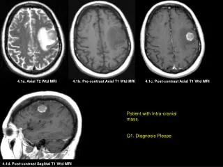
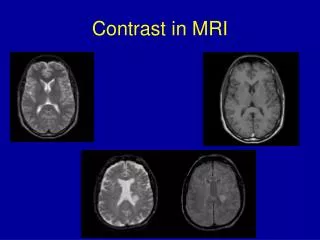
Day 1, T2 Day 6, T2 Results
Results • Using Regression Analysis, P<0.05, therefore result is significant.
Results • Using Regression Analysis, P<0.05, therefore result is significant.
Discussion • Results aren’t as expected • Paramagnetic contrast agents alter signal intensity by shortening relaxation time of protons • Increase in signal intensity is beneficial
Future Work • Contrast agent in disease diagnosis • Use this protocol on novel contrast agents
Conclusion • Results were not conclusive to show that signal intensity increased over time with the addition of a contrast agent. • More samples are needed to determine if this protocol is an effective way to test contrast agents.