Download
1 / 62
620 likes | 648 Vues
Delve into the chemical composition, structure, and functions of DNA molecules in molecular biology. Learn about nucleic acids, DNA coiling, histones, and more.

E N D
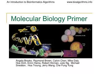
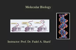
Molecular Biology (1) Mamoun Ahram, PhD Second semester, 2015-2016
Pay attention to this… • You are allowed a total absence of 15% of total credit hours (7 days) without excuses and 20% (9 days) with excuses. • If you exceed this limit, you will fail the class regardless of the 10% score. The two are independent of each other. • No points will be subtracted from your 10% for the first 3 absences.
Resources • This lecture • Campbell and Farrell’s Biochemistry, Chapter 9, 13 (pp. 381-382)
Nucleic acids • The primary structure of nucleic acids is the order of bases in the polynucleotide sequence. • The secondary structure is the three-dimensional conformation of the backbone. • The tertiary structure is specifically the supercoiling of the molecule.
Chemical composition and bonds Glycosidic bond This is why they are acidic Ribose vs. deoxyribose • Positively charged ions (Na+ or Mg2+) and peptides with positively charged side chains can associate with DNA • Eukaryotic DNA, for example, is complexed with histones, which are positively charged proteins, in the cell nucleus.
Nitrogenous bases Glycosidic bond
Nucleic acid polymer • A letter d can be added to indicate a deoxyribonucleotide residue. • for example, dG is substituted for G. • The deoxy analogue of a ribooligonucleotide would be d(GACAT).
DNA structure • Specific base-pairing • A = T; G = C; Pur = pyr • Complementary • A double helix • Backbone vs. side chains • Antiparallel • Stable • Flexible • Groovings • Stability vs. flexibility
DNA forms • B-DNA • The principal form of DNA. • Right-handed • Base pairs are perpendicular. • A-DNA • 11 base pairs per turn • Base pairs lie at an angle • Right-handed. • Wider than B-DNA • Z-DNA • Left-handed • Occurs when alternating purine–pyrimidine and sequences with methylated C • Narrower than B-DNA
DNA structure • Specific base-pairing • A = T; G = C; Pur = pyr • Complementary • A double helix • Backbone vs. side chains • Antiparallel • Stable • Flexible • Groovings • Stability vs. flexibility
DNA coiling • The bacterial circular DNA can be coiled by rotating it clockwise (negative supercoiling) or counterclockwise (positive supercoiling). • Supercoils can be added or removed by enzymes called topoisomerases: • Class I topoisomerases cut the phosphodiester backbone of one strand of DNA. • Class II topoisomerases cut both strands of DNA. • In bacteria, it is called DNA gyrase.
In eukaryotes… • In eukaryotes, DNA coiling is utilized for DNA packaging and regulation of gene activity. • Eukaryotic DNA is complexed with a number of proteins. • Chromatin = DNA molecule + proteins • The principal proteins in chromatin are the histones.
Nucleosomes • The protein core is an octamer (two molecules of histones H2A, H2B, H3, and H4). • A linker DNA/spacer region connects the octamer-DNA complexes. • A nucleosome consists of DNA wrapped around a histone core. • H1 is bound to the the octamer and wrapped DNA (a chromatosome). • Histones are positively cahrged facilitating DNA interaction and charge neutralization.
Light absorbance of nucleic acids • Aromatic pyrimidines and purines can absorb UV light • The peak absorbance is at 260 nm wavelength • The absorbance of 260 nm (A260) is constant • dsDNA: A260 of 1.0 = 50 ug/ml • ssDNA: A260 of 1.0 = 30 ug/ml • ssRNA: A260 of 1.0 =40 ug/ml Reason for ss vs. ds absorbance: • Stacked bases, vs. unstacked bases hyperchromicity What is the concentration of a double stranded DNA sample diluted at 1:10 and the A260 is 0.1? DNA concentration = 0.1 x 10 x 50 µg/ml = 50 µg /ml
Observation of denaturation • The transition temperature, or melting temperature (Tm). • Factors influencing Tm • G·C pairs • Hydrogen bonds • Base stacking • pH • Salt and ion concentration • Destabilizing agents (alkaline solutions, formamide, urea)
Central dogma of biology • Genetic information must be preserved via DNA replication. • Information must be translated into action makers (proteins) via transcription and translation. • RNA Sequence is dictated by DNA sequence.
RNA • Consist of long, unbranched chains of nucleotides joined by phosphodiester bonds between the 3’-OH of one pentose and the 5’-OH of the next • The pentose unit is β-D-ribose (it is 2-deoxy-D-ribose in DNA) • The pyrimidine bases are uracil and cytosine (they are thymine and cytosine in DNA) • In general, RNA is single stranded (DNA is double stranded).
Hybridization • DNA from different sources can form double helix as long as their sequences are compatible (hybrid DNA)
Techniques • Gel electrophoresis • Hybridization techniques
Gel electrophoresis • The length and purity ofDNA molecules can be accurately determined by the gel electrophoresis wells - - Direction DNA travels +
Resources • http://www.personal.psu.edu/pzb4/electrophoresis.swf • http://www.sumanasinc.com/webcontent/animations/content/gelelectrophoresis.html • http://www.sumanasinc.com/webcontent/animations/content/gelelectrophoresis.html
Detection Size Standard • The DNA molecules of different lengths will run as "bands“ • DNA is stained (that is, colored) with a dye (ethidium bromide) or radioactively labeled (32P) Sample 1 Sample 2 - 1000 bp 850 bp 750 bp 600 bp 200 bp 100 bp +
Hybridization techniques • Hybridization reactions can occur between any two single-stranded nucleic acid chains provided that they have complementary nucleotide sequences • Hybridization reactions are used to detect and characterize specific nucleotide sequences
Probes • A probes is a short sequence of single stranded DNA (an oligonucleotide) that is complementary to a small part of a larger DNA sequence • Hybridization reactions use labeled DNA probes to detect larger DNA fragments
Southern blotting • This technique is a combination of DNA gel electrophoresis and hybridization • Used to detect: • the presence of a DNA segment complementary to the probe • the size of the DNA fragment
1 2 1 2 1 2 : probe Southern blot Electrophoresis
Resources • This lecture • Campbell and Farrell's Biochemistry, Chapter 13, pp. 357-367; 382-384
Endonucleases • Enzymes that degrade DNA within the molecule rather than from either end (exonucleases) • Restriction endonucleases: Enzymes that recognize and cut (break) the phosphodiester bond between nucleotides at specific sequences (4- to 8-bp restriction sites) generating restriction fragments. • Type II restriction endonucleases: Always cleave always at the same place generating the same set of fragments • EcoRI (isolated from E. coli) cuts at 5'-GAATTC-3‘ • Some enzymes cut DNA at related sites • HinfI (from Haemophilus influenzae) recognizes 5'-ANTC-3' ('N' is any nucleotide) • Cuts at 5'-AATC-3', 5‘-ATTC-3', 5‘-AGTC-3' and 5'-ACTC-3’
Biological purpose of restriction endonucleases • They are present in bacteria to protect them from bacteriophages that infect bacteria by transferring their DNA into them restricting their growth.
Types of cleavages • Restriction enzymes cut DNA in two different ways: • Blunt: enzymes cut at the same position on both strands giving a blunt ended fragments • Staggered (off-center): enzymes cut the two DNA strands at different positions generating sticky or cohesive ends • The DNA fragments have short single-stranded overhangs at each end.
Palindromic sequence • The sequences recognized by restriction endonucleases—their sites of action—read the same from left to right as they do from right to left (on the complementary strand).
DNA ligase • Covalently joins DNA ends (example, restriction fragments) • Catalyzing the formation of 3’→ 5’ phosphodiester bonds between the 3-hydroxyl end of one strand and the 5-phosphate end of another strand
Advantage of restriction endonucleases • Restriction fragment length polymorphism (RFLP) • Cloning
DNA polymorphisms • Individual variations in DNA sequence may create or remove restriction-enzyme recognition sites generating different restriction fragments • Remember: our cells are diploid (alleles can be homozygous or heterozygous) • What is an allele?
Restriction fragment length polymorphism • The presence of different DNA forms in individuals generates a restriction fragment length polymorphism, or RFLP. • These can be detected by • Gel electrophoresis • Southern blotting
RFLP in the clinic • RFLP can be used as diagnostic tools. • For example, if a mutation that results in the development of a disease also causes the generation of distinctive RFLP fragments, then we can tell • if the person is diseased as a result of this mutation • from which parent this allele is inherited