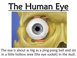
The Human Eye
A good web site for learning much about the anatomy of the human eye is : http://www.tedmontgomery.com/the_eye/. The Human Eye. Written for Physics 106 Friday, Nov. 7, 2008. Anatomy of the Human Eye. Leads to the occipital cortex at the posterior (back) of the brain .


The Human Eye
E N D
Presentation Transcript
A good web site for learning much about the anatomy of the human eye is : http://www.tedmontgomery.com/the_eye/ The Human Eye Written for Physics 106 Friday, Nov. 7, 2008
Anatomy of the Human Eye Leads to the occipital cortex at the posterior (back) of the brain
Anatomy of the Eye 3D View The greatest amount of refraction (~ 70%) occurs at the air cornea boundary Refractive index 1.38 Goes to the Brain Cataracts occur here Varies 2 to 7 mm
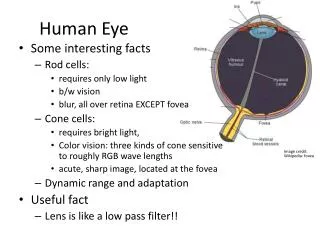
The Macula The macula lutea is the small, yellowish central portion of the retina. It is the area providing the clearest, most distinct vision. When one looks directly at something, the light from that object forms an image on one’s macula. A healthy macula ordinarily is capable of achieving at least 20/201 (“normal”) vision or visual acuity, even if this is with a correction in glasses or contact lenses. 1 Vision sharper than this sharp may be due to there being more cones per square millimeter of the macula than in the average eye, enabling that eye to distinguish much greater detail than normal.
fovea centralis The center of the macula is called the fovea centralis, an area where all of the photoreceptors are cones; there are no rods in the fovea. The fovea is the point of sharpest, most acute visual acuity. The very center of the fovea is the “foveola.” Because the fovea has no rods, small dim objects in the dark cannot be seen if one looks directly at them. For instance, to detect faint stars in the sky, one must look just to one side of them so that their light falls on a retinal area, containing numerous rods, outside of the macular zone. Rods detect dim light, as well as movement.
Why do normal vision people see color? To see any color, the retinal cone cells first must be stimulated by light. “Red-sensitive” cones are most stimulated by light in the red to yellow range, “green-sensitive” cones are maximally stimulated by light in the yellow to green range, and “blue-sensitive” cones are maximally stimulated by light in the blue to violet range. Accordingly, due to their respective sensitivities to long (L), medium (M), and short (S) wavelengths, they also are referred to as “L” cones, “M” cones, and “S” cones.
Normal Vision The process in which the lens changes its focal length to focus on objects at different distances is called accommodation
Near point and far point • The point nearest the eye at which an object can be placed and still produce a sharp image on the retina is called the near point (~ 25 cm from the eye- 20 yr old, 500 cm at age 60) • The far point of the eye is the location of the farthest object on which a fully relaxed eye can focus. Normal vision people have a far point of nearly infinity
myopia, hyperopia, astigmatism If the incoming light from a far away object focuses before it gets to the back of the eye, that eye’s refractive error is called “myopia” (nearsightedness). If incoming light from something far away has not focused by the time it reaches the back of the eye, that eye’s refractive error is “hyperopia” (farsightedness). In the case of “astigmatism,” one or more surfaces of the cornea or lens (the eye structures which focus incoming light) are not spherical (shaped like the side of a basketball) but, instead, are cylindrical or toric (shaped a bit like the side of a football). As a result, there is no distinct point of focus inside the eye but, rather, a smeared or spread-out focus. Astigmatism is the most common refractive error.
Eyeglasses for the nearsighted—A nearsighted person has a far point located only 521 cm from the eye. Assume eyeglasses ar 2 cm in front of the eye. Find the focal length needed for the diverging lenses of the glasses needed
presbyopia (“after 40” vision) After age 40, and most noticeably after age 45, the human eye is affected by presbyopia. This natural condition results in greater difficulty maintaining a clear focus at a near distance with an eye which sees clearly far away. Presbyopia is caused by a lessening of flexibility of the crystalline lens, as well as to a weakening of the ciliary muscles which control lens focusing. Both are attributable to the aging process. An eye can see clearly at a far distance naturally, or it can be made to see clearly artificially (such as with the aid of eyeglasses or contact lenses, or else following a photorefractive procedure such as LASIK). Nevertheless, presbyopia eventually will affect the near focusing of every.
Angular Magnification < 9o
Magnifying Glass Physics Remember do is related to the thin lens equation by di and f
The astronomical telescope The Refractor