Download
1 / 26
260 likes | 276 Vues
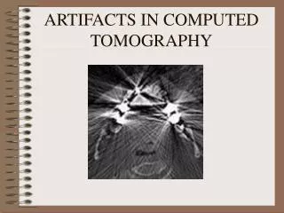
This research demonstrates how texture information from CT images can be used to classify and annotate normal tissues automatically, aiding radiologists in processing large volumes of patient data. Co-occurrence and run-length texture models are used to extract relevant features for classification.

E N D
Texture Classification of Normal Tissues in Computed Tomography 1Dong-Hui Xu, J. Lee, Daniela S. Raicu, J.D. Furst & 2David S. Channin 1Intelligent Multimedia Processing Laboratory, School of Computer Science, Telecommunications, Information Systems, DePaul University, Chicago, USA 2Department of Radiology, Northwestern University Medical School, Chicago, USA
Motivation This research will demonstrate how co-occurrence and run-length texture information from computed tomography (CT) images can be used to automatically classify and annotate normal tissues from regions of interest of heart and great vessels, liver, renal and splenic parenchyma. Automatic classification and annotation of these images will save radiologists time and assist them in processing large volumes of patient data. D. Raicu, SCAR 2005
System Diagram Input: DICOM images of Computed Tomography studies for chest & abdomen Output: Classification rules for heart, renal, splenic parenchyma, liver, and backbone D. Raicu, SCAR 2005
Segmentation • Data:340 DICOM images • Segmented organs: • liver, renal, splenic parenchyma, backbone, & heart • Segmentation algorithm:Active Contour Mappings (Snakes) • A boundary-based segmentation • algorithm with the following • inputs: • a set of initial points • five main parameters • that influence the way • the boundary is formed D. Raicu, SCAR 2005 Segmentation Segmentation
Segmentation The values of the five parameters simulate the action of two forces: Internal:designed to keep the snake smooth during the deformation External:designed to move the snake towards the boundary Output for the algorithm: The curve evolves to match the nearest internal boundary, typically based on gradient intensity measures. D. Raicu, SCAR 2005
Segmentation: Heart D. Raicu, SCAR 2005
Texture Models What is texture? Texture is a measure of the variation of the intensity of a surface, quantifying properties such as smoothness, coarseness, and regularity. Texture is a connected set of pixels satisfying a given gray level property which occurs repeatedly in an image region. D. Raicu, SCAR 2005
Texture Models Texture Models: Co-occurrence Matrix: the model captures the spatial dependence of gray-level values within an image. Texture features: entropy, variance, energy, correlation, contrast, maximum probability, homogeneity, inverse difference moment, SumMean, cluster tendency Run-Length Encoding Matrix: the model the coarseness of the texture in a specific direction. Texture features: short run emphasis (SRE) , long run emphasis (LRE), high gray-level run emphasis (HGRE), low gray-level run emphasis (LGRE), run percentage (RPC) D. Raicu, SCAR 2005
Texture Feature Extraction D. Raicu, SCAR 2005
Organ/Tissue Classification IF HGRE <= 0.38 AND CLUSTEND <= 0.048 AND INVDIFFM > 0.74 AND LRHGE > 0.46 THEN Prediction = 'Liver' Probability = 1.00 Classification rules for tissue/organs in CT images Calculate numerical texture descriptors for each region [D1, D2,…D21] D. Raicu, SCAR 2005
Organ/Tissue Classification Specifications Dataset: 66% used for training, 34% reserved for testing CART algorithm Cross-validation folds = 10 Maximum Tree Depth = 20 Parent Node/Child Node = 28/5 Minimum Change in Impurity = 0.0001 Impurity Measure = Gini Resulting Tree Total number of nodes 41 Total number of levels 8 Total number of terminal nodes 21 Resulting Rules Total number of rules: 21 (heart (3), kidneys (3), spleen (5), liver (8), and backbone (2) D. Raicu, SCAR 2005
Examples of Decision Tree Rules • IF(HGRE <= 0.38) & (CLUSTEND <= 0.05) & (INVDIFFM <= 0.74) & (SUMMEAN > 0.56) & (RLNU > 0.02) THENPrediction = ‘Renal', Probability = 0.94 • IF (HGRE <= 0.38) & (CLUSTEND > 0.05) & (SRHGE <= 0.19) & (ENTROPY <= 0.51) & (LRLGE > 0.16) THENPrediction = 'Liver', Probability = 1.00 • IF (HGRE <= 0.38) & (CLUSTEND > 0.05) & (SRHGE <= 0.19) & (ENTROPY > 0.51) & (GLNU > 0.02) THENPrediction = 'Heart', Probability = 0.96 D. Raicu, SCAR 2005
Most Significant Features • HGRE (High Gray Level Run-Emphasis) • CLUSTEND (Cluster Tendency) • HOMOGENE (Homogeneity) • INVDIFFM (Inverse Difference Moment) • SRHGE (Short Run High Gray Level Emphasis) The most important determining features for classification are located in the nodes at the top of the classification tree. D. Raicu, SCAR 2005
Classification Results Training Data D. Raicu, SCAR 2005
Classification Results Testing Data D. Raicu, SCAR 2005
Summary The results show that using only 21 texture descriptors calculated from Hounsfield unit data, it is possible to automatically classify regions of interest representing different organs or tissues in CT images. Furthermore, the results lead us to the conclusion that the incorporation of some other texture models into our proposed approach will increase the performance of the classifier, and will also extend the classification functionality to other organs. D. Raicu, SCAR 2005
Demo: HEART OPEN: To open a new Image. SEGMENT: Automatic segmentation of the regions of interest TEXTURE: Automatic calculation of the texture descriptors CLASSIFICATION: Automatic classification of the segmented regions D. Raicu, SCAR 2005
HEART: Segmentation The application allows users to change Snake / Active contour algorithm parameters D. Raicu, SCAR 2005
HEART: Segmentation (cont.) Button is clicked User selects points around the region of interest D. Raicu, SCAR 2005
HEART: Segmentation Show segmented organ If the user likes the result of the segmentation, then the user will go to the classification step D. Raicu, SCAR 2005
HEART: Classification Selection of texture models Texture features corresponding to the selected texture model are calculated and shown here D. Raicu, SCAR 2005
HEART: Classification Results are shown as follows: Predicted organ: Heart Probability: 0.86 Rule used to predict that this segmented organ is HEART D. Raicu, SCAR 2005
Following Research Projects • Project 1: Find normal tissues in CT images A. Based on segmented organs Computer –Aided Diagnosis (CAD) tools for lung cancer: • Tool 1 • Tool 2 • … heart lung backbone Goal: provide context-sensitive tools for abnormality detection & classification D. Raicu, SCAR 2005
Following Research Projects • Project 1: Find normal tissues in CT images B. Based on pure patches Goal: Develop a collection of region-of-interests (ROIs) of various tissues in normal computed tomography studies. D. Raicu, SCAR 2005
Following Research Projects • Project 2: Binning strategies for co-occurrence texture models • Linear binning • Clipped binning • Presentation will be given by Roman on linear and clipped binning C. Non-linear binning Goal: Reduce the number of gray-levels in an image such that the amount of information still present in the image will allow to differentiate among different organs/tissues D. Raicu, SCAR 2005
References • Haralick, R.M., K.Shanmugam, & I. Dinstein. Textural Features for Image Classification. IEEE Transactions on Systems, Man, and Cybernetics, vol. Smc-3, no.6, Nov. 1973. pp. 610-621. • Xu, C. & J.L. Prince. Gradient Vector Flow: A New External Force for Snakes. IEEE Proceedings of Conference on Computer Vision & Pattern Recognition, 1997. • R. Gonzalez & R. Woods. Digital Image Processing, Prentice Hall, Inc. 2002 D. Raicu, SCAR 2005