Download
1 / 71
710 likes | 810 Vues
Explore the structure of the nervous system, functions, cell types, and organization, including the CNS, PNS, afferent vs. efferent pathways, and role of glial cells. Learn about neuron anatomy and diseases like Multiple Sclerosis.

E N D
Chapter 12 – Introduction to the Nervous System Organization Cell Types
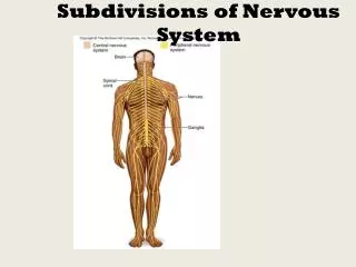
Review What 3 parts make up the nervous system? • Brain • Spinal cord • Nerves
http://www.nlm.nih.gov/medlineplus/ency/images/ency/fullsize/19588.jpghttp://www.nlm.nih.gov/medlineplus/ency/images/ency/fullsize/19588.jpg
Functions of the Nervous System • Detect changes (stimuli) in the internal or external environment • Evaluate the information • Initiate a change in muscles or glands Goal – maintain homeostasis What does this remind you of??
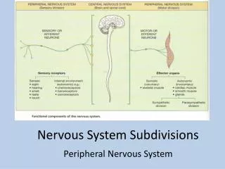
Organization of the Nervous System • Central nervous system (CNS) • Brain and spinal cord • Peripheral nervous system (PNS) • Nervous tissue in the outer regions of the nervous system • Cranial nerves: originates in the brain • Spinal nerves : originates from the spinal cord • Central fibers: extend from cell body towards the CNS • Peripheral fibers: extend from cell body away from CNS
http://www.nlm.nih.gov/medlineplus/ency/images/ency/fullsize/8679.jpghttp://www.nlm.nih.gov/medlineplus/ency/images/ency/fullsize/8679.jpg
Afferent vs Efferent Nervous pathways are organized into division based on the direction they carry information • Afferent division: incoming information (sensory) • Efferent division: outgoing information (motor) (Efferent = Exit)
Somatic & Autonomic Nervous Systems Nervous pathways are also organized according to the type of effectors (organs) they regulate • Somatic nervous system (SNS) • Somatic sensory division (afferent) • Somatic motor division (efferent)
Somatic & Autonomic Nervous Systems cont… • Autonomic nervous system (ANS): Carry information to the autonomic or visceral effectors (smooth & cardiac muscles and glands) • Visceral sensory division (afferent) • Efferent pathways • Sympathetic division – “fight or flight” • Parasympathic division – “rest and repair”
http://behavioralphys.wikispaces.com/file/view/autonomic%2520nervous%2520system.gif/162748987/autonomic%2520nervous%2520system.gifhttp://behavioralphys.wikispaces.com/file/view/autonomic%2520nervous%2520system.gif/162748987/autonomic%2520nervous%2520system.gif
Warm Up 1/5 Complete the sentences: • Afferent pathways carry… • Efferent pathways carry…. • The PNS can be subdivided into the…. • These divisions are based upon….
Review What are the two main cell types in the nervous system? (Hint: we talked about this when we covered tissue types) Answer: neurons and glia
Cells of the Nervous System Neurons: excitable cells that conduct information Glia (also neuroglia or glial cells): support cells, do not conduct information • Most numerous • Glia = glue
Types of Glia Five major types: • Astrocytes • Microglia • Ependymal cells • Oligodendrocytes • Schwann cells
Astrocytes (12-3A) • Star-shaped, largest, most numerous • Cell extension connect neurons and capillaries • Transfer nutrients from blood to neuron • Help form blood-brain barrier (BBB) http://astrocyte.info/astrocytes1.jpg
Blood-Brain Barrier • Helps maintain stable environment for normal brain function • “feet” of astrocytes wrap around capillaries in brain • Regulates passage of ions • Water, oxygen, CO2, glucose and alcohol pass freely • Important for drug research • Parkinson’s Disease
Microglia (12-3B) • Engulf and destroy cellular debris (phagocytosis) • Enlarge during times of inflammation and degeneration
Ependymal cells (12-3C) • Similar to epithelial cells • Forms thin sheets that line the fluid-filled cavities of the brain and spinal cord • Some cells help produce the fluid that fills these cavities (cerebral spinal fluid - CSF) • Cilia may be present to help circulate fluid http://www.lab.anhb.uwa.edu.au/mb140/corepages/nervous/Images/epen100he.jpg
Oligodendrocytes (12-3D) • Hold nerve fibers together • Produce myelin sheaths in CNS http://4.bp.blogspot.com/_XzEk6ORFLFg/SUQ4IitreiI/AAAAAAAAAD4/XrmtzSv1eGU/s400/article_ms_01.gif http://blustein.tripod.com/Oligodendrocytes/08-zoom.jpg
Multiple Sclerosis (MS) • Most common myelin disorder • Characterized by: • myelin loss and destruction injury and death plaque like lesions • Impaired nerve conduction weakness, loss of coordination, vision and speech problems • Remissions & relapses • Autoimmune or viral infection • Women 20-40 yrs • No known cure
Multiple Sclerosis (MS) http://www.riversideonline.com/source/images/image_popup/ww5r308_big.jpg
Schwann cells (12-3E) • Only in PNS • Support nerve fibers & form myelin sheaths • Satellite cells (12-3G) • Types of schwann cell that covers a neuron’s cell body
http://legacy.owensboro.kctcs.edu/gcaplan/anat/images/Image425.gifhttp://legacy.owensboro.kctcs.edu/gcaplan/anat/images/Image425.gif
Neurons All neurons have 3 parts: • Cell body (soma) • Axon • One or more dendrites
Neuron Anatomy • Soma resembles other cells • Nissl bodies – part of rough ER; contain proteins necessary for nerve signal transmission & nerve regeneration • Dendrites – branch out from soma; receptors; conduct impulse towards soma • Axon – process that extends from the soma at a tapered portion called the axon hillock • Axon collaterals: side branches • Telodendria: distal branches of axon • Synaptic knob: ends of telodendria
http://academic.kellogg.edu/herbrandsonc/bio201_mckinley/f14-3a_structures_in_a__c.jpghttp://academic.kellogg.edu/herbrandsonc/bio201_mckinley/f14-3a_structures_in_a__c.jpg
Neuron Anatomy • Myelin sheaths: areas of insulation produced by Schwann cells; increases speed of nerve impulse • Myelinated = white matter • Unmyelinated = gray matter • Nodes of Ravier: breaks in myelin sheath btwn Schwann cells • Synapse: junction btwn two neurons or btwn a neuron and an effector
http://academic.kellogg.edu/herbrandsonc/bio201_mckinley/f14-3a_structures_in_a__c.jpghttp://academic.kellogg.edu/herbrandsonc/bio201_mckinley/f14-3a_structures_in_a__c.jpg
Structural Classification of Neurons • Multipolar • One axon, several dendrites • Most numerous • Bipolar • One axon, one dendrite • Least numerous • Retina, inner ear, olfactory pathway • Unipolar • Axon is a single process that branches into a central process (towards CNS) and a peripheral process (towards PNS) • Dendrites at distal end of peripheral process • Always sensory neurons
http://www.google.com/imgres?imgurl=http://psyweb.com/Physiological/Neurons/NImages/Unipolarhttp://www.google.com/imgres?imgurl=http://psyweb.com/Physiological/Neurons/NImages/Unipolar http://www.google.com/imgres?imgurl=http://psyweb.com/Physiological/Neurons/NImages/multipolar http://www.google.com/imgres?imgurl=http://psyweb.com/Physiological/Neurons/NImages/bipolar
Functional Classification of Neurons • Afferent • Sensory • Towards CNS • Efferent • Motor • Towards muscles & glands • Interneurons • Connect afferent & efferent neurons • Lie within CNS
Nerves vs Tracts • Nerves – bundles of parallel neurons held together by fibrous CT in the PNS • Tracts – bundles of parallel neurons in the CNS
Warm Up 1/7 List the 5 types of glial cells and a key word/phase for each. Study for your quiz!
Warm Up 1/10 Describe the following structures: • Nissl bodies • Myelin sheaths • Axon hillock • Axon collateral • Telodendria • Synaptic knobs
Examples of Reflex Arcs • Ipsilateral • Contralateral • intersegmental
Nerve Fibers • Remember the difference between nerves and tracts? • Endoneurium: surrounds each nerve fiber • Perineurium: surrounds fascicles (bundles of nerve fibers • Epineurium: surrounds a complete nerve (PNS) or tract (CNS)
Review: Gray vs White Matter • White matter – myelinated nerve fibers • Myelin sheaths help increase the speed of an action potential • Gray matter – unmyelinated nerve fibers & cell bodies • Ganglia: regions of gray matter in PNS
Nerve Fiber Repair • Nervous tissue has a limited repair capacity b/c mature neurons are incapable of cell division • Repair can take place if soma and neurilemma remain intact
Steps of Nerve Fiber Repair • Injury • Distal axon and myelin sheaths degenerates • Remaining neurilemma & endoneurium forms a “tunnel” from the injury to the effector • Proteins produced in the nissl bodies help extend a new axon down the tunnel to the effector
Nerve Impulses • Neurons are specialized to initiate and conduct signals nerve impulses • Exhibit excitability & conductivity • Nerve impulse wave of electrical fluctuation that travels along the plasma membrane
Membrane Potentials • Difference in charges across the plasma membrane • Inside slightly negative • Outside slightly positive • Result in a difference in electrical charges membrane potential • Stored potential energy • Analogy = water behind a dam
Membrane Potentials • Membrane potential creates a polarized membrane • Membrane as – pole & + pole • Potential difference of a polarized membrane is measured in millivolts (mV) • The sign indicates the charge of the inside of a polarized membrane
Resting Membrane Potential (RMP) • When not conducting electrical signals, a membrane is “resting” • -70mV • RMP maintained by ionic imbalance across membrane • Sodium-Potassium Pump • Pumps 3 Na+ out for every 2 K+ pumps in • Creates an electrical gradient (more positive on outside)
Local Potential • Local potential - The slight shift away from the RMP • Isolated to a particular region of the plasma membrane • Stimulus-gated Na+ channels open Na+ enters membrane potential to moves closer to zero (depolarization) • Stimulus-gated K+ channels open K+ exits membrane potential away from zero (hyperpolarization) • **Local potentials do not spread to the end of the axon**