Investigating the Impact of PTB Knockdown on BAG-1 Isoform Expression in HeLa Cells
This study examines the effect of PTB knockdown on BAG-1 isoform expression in HeLa cells. Cells were transfected with siRNAs targeting PTB and a control sequence, followed by transfection with either the di-cistronic plasmid pRBagF or control plasmid pRF. Luciferase activity was measured using a Dual-Luciferase reporter assay system, and Western analysis was performed to assess PTB and BAG-1 expression levels, with actin as a loading control. Results indicate a significant reduction in BAG-1 p36 isoform levels and IRES activity upon PTB knockdown.

Investigating the Impact of PTB Knockdown on BAG-1 Isoform Expression in HeLa Cells
E N D
Presentation Transcript
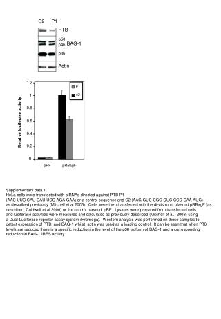
C2 P1 PTB p50 BAG-1 p46 p36 Actin 1.2 1 0.8 p1 0.6 Relative luciferase activity c2 0.4 0.2 0 pRF pRBagF Supplementary data 1. HeLa cells were transfected with siRNAs directed against PTB P1 (AAC UUC CAU CAU UCC AGA GAA) or a control sequence and C2 (AAG GUC CGG CUC CCC CAA AUG) as described previously (Mitchell et al 2005). Cells were then transfected with the di-cistronic plasmid pRBagF (as described; Coldwell et al 2000) or the control plasmid pRF. Lysates were prepared from transfected cells and luciferase activities were measured and calculated as previously described (Mitchell et al., 2003) using a Dual-Luciferase reporter assay system (Promega). Western analysis was performed on these samples to detect expression of PTB, and BAG-1 whilst actin was used as a loading control. It can be seen that when PTB levels are reduced there is a specific reduction in the level of the p36 isoform of BAG-1 and a corresponding reduction in BAG-1 IRES activity.
![Altitude= [0.2, 4.0km]](https://cdn4.slideserve.com/464372/nasa-s-cloud-absorption-radiometer-dt.jpg)