
Systemic Immune Complex Disease
E N D
Presentation Transcript
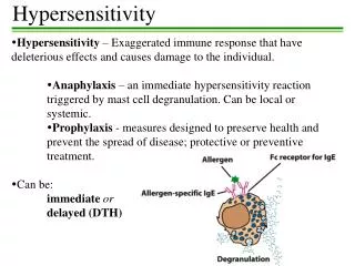
Type III Hypersensitivity (ImmuneComplex - Mediated) • Type III hypersensitivity is mediated by the deposition of antigen-antibody (immune) complexes, followed by complement activation and accumulation of polymorphonuclear leukocytes. • Immune complexes can involve exogenous antigens such as bacteria and viruses or endogenous antigens such as DNA.
Pathogenic immune complexes either form in the circulation and subsequently deposit in the tissues or form at extravascular sites where antigen has been planted (in situ immune complexes).
Immune complex-mediated injury can be systemic when complexes are formed in the circulation and are deposited in multiple organs or localized to particular organs (e.g. kidneys, joints, or skin) if the complexes are formed and deposited in a specific site.
SYSTEMIC IMMUNE COMPLEX DISEASE • The pathogenesis - three phases: • (1) formation of antigen antibody complexes in the circulation (2) deposition of the immune complexes in various tissues, (3) inflammatory reaction in various sites throughout the body. • Acute serum sickness is the prototype of a systemic immune complex disease.
Approximately 5 days after a foreign protein is injected, specific antibodies are produced; these react with the antigen still present in the circulation to form antigen-antibody complexes (first phase). In the second phase, antigen-antibody complexes formed in the circulation deposited in various tissue beds
SYSTEMIC IMMUNE COMPLEX DISEASE • Two important factors determine whether immune complex formation leads to tissue deposition and disease: • Size of the immune complex: Very large complexes formed in antibody excess are rapidly removed from the circulation by the mononuclear phagocytic cells and are therefore' relatively harmless. • The most pathogenic complexes are formed during antigen excess and are small or of intermediate size, are cleared less avidly by phagocytic cells, and therefore circulate longer.
Status of the mononuclear phagocyte system: Because macrophages normally filter out circulating immune complexes their overload or dysfunction leads to persistence of immune complexes in the circulation and increases the probability of tissue deposition.
SYSTEMIC IMMUNE COMPLEX DISEASE • In addition, several other factors influence whether and where immune complexes deposit. • These include charge of the complex (anionic vs. cationic), valency of the antigen, avidity of the antibody, affinity of the antigen for various tissues, three-dimensional architecture of the complexes, the hemodynamics of a given vascular bed.
The favored sites of immune complex deposition are -kidney, joints, skin, heart, serosal surfaces and small blood vessels. • Localization in the kidney is explained in part by the filtration function of the glomerulus, with trapping of the circulating complexes in the glomeruli.
SYSTEMIC IMMUNE COMPLEX DISEASE • For complexes to leave the circulation and deposit within or outside the vessel wall, an increase in vascular permeability must occur. • This is probably mediated when immune complexes bind to inflammatory cells via Fc and C3b receptors and trigger release of vasoactive mediators and/or permeability-increasing cytokines.
Once complexes are deposited in the tissue, the third phase, inflammatory reaction, ensues. • During this phase (approximately 10 days after antigen administration), clinical features such as fever, urticaria, arthralgias, lymph node enlargement, and proteinuria appear.
SYSTEMIC IMMUNE COMPLEX DISEASE • Tissue damage is also mediated by oxygen free radicals produced by activated neutrophils. • Complement activation by immune complexes is central to the pathogenesis of injury, releasing biologically active fragments such as the anaphylatoxins ( C3a and C5a), which increase vascular permeability and are chemotactic for polymorphonuclear leukocytes.
Phagocytosis of immune complexes by the accumulated neutrophils results in the release or generation of a variety of additional proinflammatory substances, including - prostaglandins, vasodilator peptides, and chemotactic substances lysosomal enzymes capable of digesting basement membrane, collagen, elastin, and cartilage.
Tissue damage is also mediated by oxygen free radicals produced by activated neutrophils
SYSTEMIC IMMUNE COMPLEX DISEASE • Immune complexes can also cause platelet aggregation and activate Hageman factor; both of these reactions augment the inflammatory process initiate microthrombi formation • that contribute to the tissue injury by producing local ischemia. • The resultant pathologic lesion is termed vasculitis if it occurs in blood vessels, glomerulonephritis if it occurs in renal glomeruli, arthritis if it occurs in the joints.
Obviously, only complement-fixing antibodies (i.e., IgG and IgM) can induce such lesions. • Because IgA can also activate complement by the alternative pathway, IgA-containing complexes may also induce tissue injury. • During the active phase of the disease, consumption of complement decreases the serum levels.
LOCAL IMMUNE COMPLEX DISEASE(ARTHUS REACTION) • The Arthus reaction may be defined as a localized area of tissue necrosis resulting from acute immune complex vasculitis. • The reaction is produced experimentally by injecting an antigen into the skin of a previously immunized animal (i.e., preformed antibodies against the antigen are already present in the circulation).
Because of the initial antibody excess, immune complexes are formed as the antigen diffuses into the vascular wall; these are precipitated at the site of injection and trigger the same inflammatory reaction
Arthus lesions evolve over a few hours and reach a peak 4 to 10 hours after injection, when the injection site develops visible edema with severe hemorrhage occasionally followed by ulceration.
Type IV Hypersensitivity (Cell-Mediated) • Cell-mediated immunity is the principal mechanism of response to a variety of microbes, including intracellular pathogens such as Mycobacterium tuberculosis and viruses, as well as extracellular agents such as- fungi, protozoa, and parasites.
However, these processes can also lead to cell death and tissue injury, either as a consequence of the normal clearance of infection or in response to self-antigens (in autoimmune disease). • So-called contact skin sensitivity to chemical agents and graft rejection are other instances of cell-mediated hypersensitivity reactions.
Type IV Hypersensitivity (Cell-Mediated) • Type IV hypersensitivity is mediated by specifically sensitized T cells rather than by antibodies and is subdivided into two basic types:- (I) delayed-type hypersensitivity, initiated by CD4+ T cells, (2) direct cell cytotoxicity, mediated by CD8+ T cells.
In delayed hypersensitivity, TH1-type CD4+ T cells secrete cytokines, leading to recruitment of other cells, especially macrophages, which are the major effector cells. • In cell-mediated cytotoxicity, cytotoxic CD8+ T cells assume the effector function.
Delayed hypersensitivity (DTH) • A classic example - Tuberculin reaction, elicited in an individual already sensitized to the tubercle bacillus by a previous infection. • Eight to 12 hours after intracutaneous injection of tuberculin a local area of erythema and induration appears reaching a peak (typically 1 to 2 cm diameter) in 24 to 72 hours (hence the adjective, delayed) and thereafter slowly subsiding.
Histologically, the DTH reaction is characterized by the perivascular accumulation ("cuffing") of CD4+ helper T cells and, to a lesser extent, macrophages.
Delayed hypersensitivity (DTH) • Local secretion of cytokines by these mononuclear inflammatory cells leads to an associated increased microvascular permeability, giving rise to dermal edema and fibrin deposition; the latter is the main cause of the tissue induration in these response.
The tuberculin response is used to screen populations for individuals who have had prior exposure to tuberculosis and therefore have circulating memory T cells. Notably, immunosuppression or loss of CD4+ T cells (e.g., owing to HIV) may lead to a negative tuberculin response even in the presence of a severe infection.
Delayed hypersensitivity (DTH) • The sequence of events in DTH (as exemplified by the tuberculin reaction) begins with the first exposure of the individual to tubercle bacilli. • CD4+ lymphocytes recognize peptide antigens of tubercle bacilli in association with class II antigens on the surface of monocytes or dendritic cells that have processed the mycobacterial antigens. • This process leads to the formation of sensitized CD4+ cells of the TH1 type that remain in the circulation for years.
On subsequent cutaneous injection of tuberculin into such an individual, the memory cells respond to processed antigen on APCs and are activated (undergo blast transformation and proliferation), accompanied by the secretion of T H1 cytokines. • It is these T H1 cytokines that are ultimately responsible for driving the development of the DTH response
Delayed hypersensitivity (DTH) The most relevant cytokines in the process include the following: • IL-12 is a cytokine produced by macrophages after initial interaction with the tubercle bacillus. • It is critical for the induction of DTH in that it is the major cytokine that drives the differentiation of TH1 cells. • IL- 12 is also a potent inducer of IFNγ secretion by T cells and NK cells
Delayed hypersensitivity (DTH) IFN γ • It is an extremely potent activator of macrophages increasing macrophage production of IL-12. • Activated macrophages express more class II molecules on the surface, leading to augmented antigen presentation capacity. • .
They also have increased phagocytic and microbicidal activity, and their capacity to kill tumor cells is enhanced. • Activated macrophages secrete several polypeptide growth factors, including platelet derived growth factor (PDGF) and TGF-α, which stimulate fibroblast proliferation and augment collagen synthesis.
Delayed hypersensitivity (DTH) • IL-2 causes the proliferation of the T cells that have accumulated at sites of DTH. • TNF and lymphotoxin are cytokines that exert important effects on endothelial cells: (1) increased secretion of nitric oxide and prostacyclin, favoring increased blood flow via local vasodilation; (2) increased expression of E-selectin , an adhesion molecule promoting mononuclear cell attachment; (3) induction and secretion of chemotactic factors such as IL-8.
GRANULOMATOUS INFLAMMATION • A granuloma is a special form of DTH occurring in the setting of persistent and/or nondegradable antigens. • The initial perivascular CD4+ T-cell infiltrate is progressively replaced by macrophages over a period of 2 to 3 weeks; these accumulated macrophages typically exhibit morphologic evidence of activation, that is, they become large, flat, and eosinophilic (denoted as epithelioid cells).
A granuloma is a special form of DTH occurring in the setting of persistent and/or nondegradable antigens. • The initial perivascular CD4+ T-cell infiltrate is progressively replaced by macrophages over a period of 2 to 3 weeks; these accumulated macrophages typically exhibit morphologic evidence of activation, that is, they become large, flat, and eosinophilic (denoted as epithelioid cells).
The epithelioid cells occasionally fuse under the influence of certain cytokines (e.g., IFN γ) to form multinucleated giant cells. • A microscopic aggregate of epithelioid cells, typically surrounded by a collar of lymphocytes, is called a granuloma, and the pattern is referred to as granulomatous inflammation.
Delayed hypersensitivity (DTH) • DTH is a major mechanism of defense against a variety of intracellular pathogens, including mycobacteria, fungi, and certain parasites and it may also be involved in transplant rejection and tumor immunity. • The central role of CD4+ T cells in delayed hypersensitivity is evident in patients with AIDS.
Because of the loss of CD4+ cells, the host response against intracellular pathogens such as Mycobacterium tuberculosis is markedly impaired. • The bacteria are engulfed by macrophages but are not killed, and instead of granuloma formation,there is accumulation of unactivated macrophages poorly adapted to deal with the invading microbe.
Delayed hypersensitivity (DTH) • In addition to its beneficial, protective role, DTH can also be a cause of disease. • Contact dermatitis is one such example of tissue injury resulting from delayed hypersensitivity. • It is evoked by contact with pentadecylcatechol in a sensitized host and manifests as a vesicular dermatitis.
On re-exposure to the plants, sensitized TH1 CD4+ cells accumulate in the dermis and migrate toward the antigen within the epidermis. • Here they release cytokines that damage keratinocytes, causing separation of these cells and formation of an intraepidermal vesicle
T-CELL - MEDIATED CYTOTOXICITY • In this form of type IV hypersensitivity, sensitized CD8 + T cells kill antigen-bearing target cells. • Class I MHC molecules bind to intracellular viral peptides and present them to CD8+ T lymphocytes. • The CD8+ effector cells, called cytotoxic T lymphocytes(CTLs), play a critical role in resistance to virus infections.
The lysis of infected cells before viral replication is completed leads ultimately to elimination of the infection. • It is believed that many tumor-associated peptide are also presented on tumor cell surfaces, and hence CTLs may also be involved in tumor immunity.
T-CELL - MEDIATED CYTOTOXICITY • Two principal mechanisms of CTL killing have been demonstrated (1) perforin-granzyme-dependent killing (2) Fas-Fas ligand-dependent killing. • Perforins and granzymes are soluble mediators contained in the lysosome- like granules of CTLs. • Perforin punches holes in the plasma membrane of target cells; it does so by insertion and polymerization of perforin molecules to form a pore.
These pores allow water to enter the cells, eventually resulting in osmotic lysis. • The lymphocyte granules also contain a variety of proteases called granzymes, which are delivered into the target cells via the perforin pores. • Once inside the cell, granzymes activate target cell apoptosis.
T-CELL - MEDIATED CYTOTOXICITY • Activated CTLs also express Fas ligand (a molecule with homology to TNF), which binds to Fas on target cells. • This interaction leads to apoptosis. • In addition to viral and tumor immunity, CTLs directed against cell surface histocompatibility antigens also play an important role in graft rejection.