Download
1 / 30
310 likes | 323 Vues
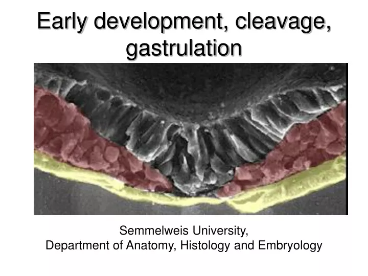
Earl y development , c leavage , gastrulatio n. Semmelweis University , Department of Anatomy, Histology and Embryology. The zygote =fertilized egg. The new diploid cell is created by the fusion of the male and female pronucleus ( ). Cleavage. Mitotic division process

E N D
Early development, cleavage, gastrulation Semmelweis University, Department of Anatomy, Histology and Embryology
The zygote =fertilized egg The newdiploidcell is createdbythefusion of themale and femalepronucleus ( )
Cleavage • Mitotic division process • Cleavage stage cells: blastomeres • The volume of egg’s cytoplasm divides into numerous smaller, nucleated cells → in humans whole cytoplasmatic volume of the egg packs into smaller and smaller blastomeres • The human cleavage is equal, but asynchronous
Different type of cleavage • The cleavage process is influenced by the yolk contents of the egg. • The egg may contain large or small amount of yolk • The distribution of the yolk: • uniformly • concentrate on one of the pole of the egg. • Due to the asymmetric yolk distribution: • Only a part of the egg pack into blastomeres • blastomeres have different size • Complete and incomplete types of cleavage can be distinguished Incomplete (Meroblasticus) fish, reptiles, birds Frog Complete (Holoblasticus) Human
What stage is DNA in embryo first transcribed? Little increase in RNA during the first 2-8 days. In the mouse as early as 2 cell, by blastocyst stage produces adult levels. Protein synthesis occurs at 2-4 cell stage where there is a large increase. In most species, the rate of cell division and the placement of the blastomeres are completely under the control of the proteins and mRNAs that are stored in the oocyte cytoplasma. So they come from only the mother. The zygotic genome does not function in early-cleavage embryos until 2-4 cells stage in human. Because the sperm and egg contains highly compressed and methylated DNA which are not capable to be transcripted. After the 4 cell stage, the embryonic DNA gradually demethylated and transcription of proteins starts from it. Evidence indicates that maternal and embryonic mRNA are used during early cleavage, with loss of maternal RNA by blastocyst stage. If you block RNA synthesis with actinomycin D (reduces RNA synthesis by 90%) and depress cleavage, only reduces protein synthesis by 50%. Evidence indicates that the embryonic genome does not begin to control the course of development until blastulation.
Compaction -in the 8-cell-stage cleavage. -The blastomeres, which show loose arrangement, suddenly form a compact ball -Before the compaction blastomeres loosly adhere to each other by microvilli on their surface. -After compaction blastomeres tightly adhere with each other through intercellular junctions before compaction and after compaction
Compaction is a membrane polarization process → well-defined apical, basal and lateral side is developing. • Different components of the cell membrane concentrate at different regions of the cell causing the polarization of the cells. • Membrane polarization is influenced by cell-cell interactions • This polarization process takes place only in those parts of the cell membrane where the cell is in contact with other blastomeres. • E-cadherin plays a main role in compaction. • At 2-cell stage, E-cadherin is uniformly spread throughout the cell membrane. During compaction E-cadherin becomes restricted to those sites of cell membrane where adjacent blastomeres are in contact with each other. • E-candherin molecules accumulate and form zonula adherens
Blastula formation • Newlyformedstructuresbetweentheouterblastomeres: • -tightjunctions (apical part) • -gapjuction(lateral part) • -membranetransportmoleculesonbasal part: mainlysodiumpumps. • Duetotheactivatedsodiumpumpsthesodiumconcentrationincreaseintheintercellularspace and parallel thewaterflowsintotheintercelullarspacebyosmosis → forming fluid filledcavitycalledblastocoel. • The blastocoel is expandedgraduallybytheincreasingamount of the fluid
The expanding blastocoel push the internal cells on the one side of the blastocyst → inner cell mass (ICM) In mice: • trophoblast cells differentiate into the fetal membrane system • inner cell mass forms the whole body of the embryo and extraembryonic edoderm
Twins • Monozygotictwinsarisefromonezygote • Dizygotetwinsarisetwodifferenteggs, thatare fertilised bytwodifferentsperms • .Monozygotictwins • Atcleavage:afterthefirstcelldivisionthenewlyformedtwoblastomerescompletelyseparetefromeachother. • Atblastulaion: thesubdivision of theinnercellmasswithintheblastocyst.
The differentiation of the ICM cellsinto a bilaminarstructurecontainingepiblast and hypoblastlayer. The sortingmodel. Atthebeginning, theepiblast and hypoblastcellsdistributewithinthe ICM showing „salt and pepperpattern”. The sortingevent, inwhichthebilaminardiscwill be developed, is causedbytwo main ways. 1.: thedifferentstrenghts of adhesionbetweenthetwocellstype (adhesion is strongerbetweensametypecellsthandifferenttypecells) and 2.: signalscomingfromeithertheblastocoelorfromtrophoblast (theepiblastcellsexpressnanogwhilehypoblastcellsexpressGATA6 and thisexpressionpattern is causedbythedifferentFGF signaling.
Why is gastrulation so important? • Generation of the basic body plan. • Specification of the axes: • Anterior and posterior • Dorsal and ventral • Left and right • Generation of the three germ layers • Ectoderm, mesoderm, and endoderm
Primitivestreak, groove Anterior Posterior • Gastrulation begins with the formation of primitive streak. Epiblast ploriferate and they are pushed toward the midline of the embryo → they are jammed in the midline forming the primitív streak that first appear in the posterior part of the embryo. • Cells ,which are located in the middle of the primitív streak, migrate into the interior of the embryo resulting the formation of primitív groove in the middle of the primitív streak.
Hensen’s node • The primitív streak with the primitív groove is growing gradually anteriorly • At the anterior end of the primitív streak there is a small but well-defined accumulation of cells, called primitív node or Hensen’s node. cranial side caudal side Cranio-caudal, left-rightaxeswell-defined!
What ‘s happen with the cells in the primitive groove? • The movements of the cells are accopamanied by major changes in their structure. • When epiblast cells enter the primitív streak, they become elongated and lose their connection with their adjacent cells → their morphologes change forming bottle cells. • Within the primitív groove these bottle cells lose their connection with the basal lamina become free from the epiblast layer. • Bottle cells undergo an epithelio-mesenchymel transformation within the primitív groove and the newly form mesenchymel cells are able to migrate as individual cells. • During the epithelio-mesenchymal transformation the E-cadherin synthesis downregulate within the bottle cells, Epithelial cell E-cadherin epiblast „slug” FGF8 E-cadherin mesoderm endoderm
Development of the definitive endoderm D14-15 Bi-laminar embryonic disk Hypoblast cells develop only into extraembryonal mesoderm Primitive streak Epiblast Hypoblast First entering epiblast-cells migrate and replace the hypoblast-cells forming the definitive endoderm Endoderm Epiblastcellsgiverisetothethreegermlayers of theembryo!!
Development of the Intraembryonal Mesoderm Primitive streak D16 epithelio-mesenchymal transformation Intraembryonal mesoderm Definitive entoderm Epiblast-cells migrate in the interlaminar space and forming intraembryonal mesoderm
Migration of the Mesodermal Cells • The newly form mesenchymal cells migrate and spread bilaterally. • Those cells, which pass through at the level of Hensen’s node, migrate directly cranially and form the precordal plate and later take part in the formation of the notochord. Epiblast Mesoderm Entoderm Epiblast Entoderm
Primitiv streak regression • At the beginning of gastrulation the primitiv streak grows cranially • The primitiv streak growing changes for its regression. → its length is decreasing toward caudally • This regression process is related with the elongatoin of the notochord • The notochord is formed by the addition of cells to its caudal end while the primitiv streak become shorter and shorter
1.: At the biginnig of the notochord process first form a notochord canal with a central lumen Human notochord 3.:The notochord plate starts to infold later form the rope like notochord 2.:The floor of the notochordal canal disappears remaining a flatten plate (notochordal plate) which incorporate into the definitiv endoderm
Differentation of the mesoderm, convergent extension membrana buccopharyngea paraxial mesoderm (somites) intermediate mesoderm (nephrotom) lateral mesoderm (somatopleura, splanchnopleura) primitive node primitive strike extraembryonal mesoderm
Fate map GE: gut endoderm PP: prechordal platePS: primitive streak CM: cardiac mesodermPEEM: extraembryonic mesoderm HM: head mesoderm S: somitic mesoderm IM: intermedier mesoderm LPM: lateral plate mesoderm SE: surface ectoderm NP: neural plate PE: placod ectoderm NC: neural crest
Gastrulation wall of amnion roof of blastocyst extra- embryonic mesoderm hypoblast epiblast bi-laminar mesoblast (mesenchyma) embryonic ectoderm embryonic entoderm three-laminar intraembryonic mesoderm primary mesoderm definitive entoderm definitive ectoderm Connective tissue, vessels, smooth and skeletal muscle, blood cells, skeleton, genito-urinary organs lining epithelium and the glands of the respiratory and gastrointestinal organs skin, nervous system, sensory organs