Download
1 / 16
160 likes | 352 Vues
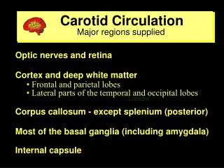
Carotid Circulation Major regions supplied . Optic nerves and retina Cortex and deep white matter Frontal and parietal lobes Lateral parts of the temporal and occipital lobes Corpus callosum - except splenium (posterior)

E N D
Carotid Circulation Major regions supplied • Optic nerves and retina • Cortex and deep white matter • Frontal and parietal lobes • Lateral parts of the temporal and occipital lobes • Corpus callosum - except splenium (posterior) • Most of the basal ganglia (including amygdala) • Internal capsule ventricle Lumen
ACA Key functional areas supplied • Septal area • Primary motor cortex • Leg and foot areas • Area controlling urinary bladder • Motor planning areas in medial frontal lobe, anterior to the precentral gyrus • Primary somatosensory cortex for leg and foot • Most of corpus callosum except splenium ventricle Lumen
MCA-superior branches Key functional areas supplied • Primary motor cortex - face / arm areas • Pyramidal tract axons from all of the primary • motor cortex • Primary somatosensory cortex - face / arm areas • Frontal eye fields • Frontal/parietal - language-dominant hemisphere • Broca’s and other areas for language expression • Frontal/parietal - nondominant hemisphere • areas for 3-D visuospatial concepts of self and world • areas for expressing emotions ventricle Lumen
MCA-inferior branches Key functional areas supplied • Visual radiations • Fibers representing information from contralateral • superior quadrants • Temporal/parietal- language-dominant hemisphere • Wernicke’s and other areas for language comprehension • Posterior parietal - nondominant hemisphere • areas for 3-D visuospatial perceptions of self and world • areas for interpreting emotions ventricle Lumen
Vertebral-Basilar Circulation Major regions supplied • Upper cervical spinal cord • Brainstem and cerebellum • Most of thalamus and hypothalamus • Cortex and deep white matter • Posterior medial parietal lobes • Medial and infrior temporal and occipital lobes • Splenium of corpus callosum ventricle Lumen
PCA Key functional areas supplied • By long penetrating branches • Thalamus, subthalamic nucleus, hypothalamus • Midbrain • By cortical branches - parietal and occipitallobes • Visual radiations and primary visual cortex • Splenium of corpus callosum • By cortical branches - medial temporal lobe • Hippocampal formation and posterior fornix ventricle Lumen
Common Anastomoses Collateral circulation may prevent ischemia • External-internal carotid via ophthalmic branches • Circle of Willis • Muscular branches of cervical arteries-vertebral • or external carotid arteries • Short cortical branches of ACA, MCA, PCA • Branches of the major cerebellar arteries ventricle Lumen
ACA-MCA Border Zone Key functional areas that may be present • Hip or shoulder (occasionally arm) regions of primary motor or somatosensory cortex • Regions involved in aspects of language production (in the dominant hemisphere) • Frontal Eye Fields • Motor planning areas in frontal lobe ventricle Lumen
ACA/MCA-PCA Border Zone Key functional areas that may be present Visual radiations Foveal representation in the primary visual cortex Inferior temporal lobe cortex for visual recognition Regions for language comprehension and word finding (possibly reading) in dominant hemisphere Regions important for visuospatial perceptions in parietal lobe of nondominant hemisphere ventricle Lumen
What is a TIA? A Transient Ischemic Attack (TIA) is a brief episode in which neurologic deficits suddenly occur, but then disappear completely • Most TIAs last a few minutes to an hour. • No neurologic deficits remain once a TIA • has ended, because little or no brain tissue is • permanently damaged. • A TIA is an indicator that the stage is set • for an ischemic stroke. • Treatment of patients with TIAs can significantly • reduce their risk of having a stroke. ventricle Lumen
TIAs: Carotid Territory Transient monocular blindness TMB (also known as amaurosis fugax) occurs when the retina becomes temporarily ischemic. • Patients commonly describe a gray or black fog • or a mist clouding vision in all or part of one eye. • Attacks are typically brief (1-5 minutes), and • afterwards vision is fully restored. • TMB often signals the presence of severe • ipsilateral carotid artery disease in older adults. • TMB can also be caused by migraine.
TMB: What’s Mechanism? • Two Ideas: • 1. Thrombus from ulcerated atherosclerotic plaque at the origin of the internal carotid artery enters ophthalmic artery and plugs retinal vessel. • A small embolic particle can produce retinal ischemia • or block the central retinal artery. • If the embolus (often a platelet-fibrin aggregate) falls • apart, blood flow is restored and and vision returns. • 2. Low perfusion in the internal carotid artery, putting distal structures like the retina at risk. • This mechanism implies presence of severe carotid • stenosis that significantly impairs blood flow.
TIAs: Carotid Territory Transient hemispheric attacks Problems typically produced • One-sided limb weakness, clumsiness or paralysis • One-sided numbness, paresthesia, or sensory loss • Difficulty with language production or • comprehension • Inability to articulate words clearly, often • described as ‘I slurred my words’ (dysarthria) • Partial or complete homonymous visual field • defects (patients seldom describe these)
Carotid Bifurcation atherosclerotic plaque and thrombus Thrombus on plaque surface Plaque Thrombus on plaque surface Plaque reduces the diameter of artery
TIAs: Vertebral-Basilar Various combinations may be present Problems typically produced • Vertigo or dizziness • Unilateral or bilateral weakness or clumsiness • Unilateral or bilateral numbness or sensory loss • Limb ataxia or coarse tremor, staggering gait • Dysarthria • Visual field defect, blindness, or diplopia • Nystagmus (‘it jumps around when I look at it’)
Patients may ignore TIAs! • Because the episode of impaired function is brief, patients may not tell you about it unless you ask. • Seniors with somewhat impaired memory may simply not recall such brief events. • TIAs in carotid territory predict severe atherosclerosis • in the proximal internal carotid artery. • Tests can determine actual blood flow in the carotid. • The odds of stroke in the next 1-2 weeks are great! • If severe narrowing is present, surgical and medical • treatments can help to reduce stroke risk.