Degloving Injuries
1.08k likes | 7.56k Vues
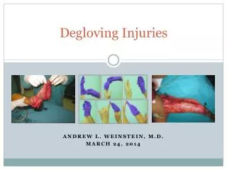
Degloving Injuries. Andrew L. Weinstein, M.D. March 24, 2014. What is a degloving injury?. A type of avulsion in which an extensive section of skin is completely torn off the underlying tissue, severing its blood supply So-called for its resemblance to removing a glove.

Degloving Injuries
E N D
Presentation Transcript
Degloving Injuries Andrew L. Weinstein, M.D. March 24, 2014
What is a degloving injury? A type of avulsion in which an extensive section of skin is completely torn off the underlying tissue, severing its blood supply So-called for its resemblance to removing a glove
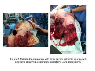
Initial evaluation • Great force (MVC, industrial, agricultural) serious co-injury to other organ systems • Treat life-threatening injuries before proceeding to evaluation and treatment of extremity injury, which itself is rarely life-threatening • ATLS • Primary survey • Airway maintenance with cervical spine protection • Breathing and ventilation • Circulation with hemorrhage control • Disability/neurologic assessment • Exposure and environmental control • Secondary survey
Initial evaluation: “when, where, how” • When: • Longer duration = greater risk for infection (>6h precludes primary closure or immediate coverage) • Sensitivity to ischemia (muscle, 4-6h > skin > bone) • Temperature (12-24h viability of devascularized tissues if cooled) • Where • Farming = highly contaminated (aggressive debridement, precludes primary closure or immediate coverage) • How • Force of injury = extent of tissue necrosis or “zone of injury” (aggressiveness of approach, removal of foreign bodies, compartment syndrome)
Physical exam and work-up Vital signs HPI: wound contamination/tetanus status Physical exam: neurovascular, musculoskeletal Plain films, scans Labs: CBC, BMP (K), T&S, CK, UA, EKG
A note about capillary refill and nerve findings • Nail bed capillary refill unreliable indicator of peripheral perfusion (stagnant blood in devascularizeddigit) • More reliable is dorsal paronychial tissue on side of nail • Most reliable indicator is color of blood that oozes from tissue after needle prick (bright red vs. purple) • For major vessels, use handheld Doppler probe • Nerve can remain physically intact after crush/avulsion injury, yet axons may still be damaged • Neuropraxia vs. axonotmesis
Initial debridement • Single most important step • If inadequate and nonviable tissue left behind • Infection • Further tissue loss • Potential loss of limb or life • If skin does not bleed or oozes only dark blood at the time of initial surgery, must debride to create healthy soft tissue bed for reconstruction • If wound is heavily contaminated or involves critical areas where viability uncertain, repeat OR debridement 24-48h later
Initial debridement cont’d… Although early wound closure desirable, often prudent to delay definitive coverage until wound stable (e.g. reduced contamination) Serial debridement separated by 24-48h to reduce infection and optimize healing and motion Aim for definitive coverage by 7-10d with in interim keeping vital structures moist As swelling develops after injury, tendency for wounds to enlarge making closure more difficult
Wound reduction • “Vessel loops” • Crisscross fashion with staples along wound edge to create “corset effect” • Brings wound together without ischemia, avoids compartment syndrome • NPT/VAC = “mechanical fibroblast” • Removal of exudate, decrease in edema, closure of dead space, promotion of wound contraction, and promotion of granulation • Wound contracts and granulates, covered with skin graft rather than complex flap
VAC: special considerations • Closed system change sponge q3-5d or more frequently depending on level of contamination • Revolutionized approach to soft tissue coverage in complex lower extremity defects • Should NOT be used for extremities with • Severe contamination • Infection • Significant bleeding • Caution in setting of vascular repair or reconstruction
Soft tissue coverage Goal: achieve healed wound with stable, durable coverage and vascularized tissue over critical structures Determines environment in which all other repaired and reconstructed structures will heal and function Coverage should be low profile and supple over mobile areas such as joints
Reconstruction • The simplest method of coverage appropriate to the situation to achieve optimum form and function • Secondary intention • Primary closure • Skin grafting • STSG • FTSG • NPT/VAC • Flap • Local • Distant • Free
Additional considerations for surgery • Patients health • Age, CV/pulmonary disease, bleeding tendencies, DM increase risk of perioperative complication or even mortality • Consider simplifying the method of reconstruction • Smoking or use of vasoactive drugs (e.g. cocaine) is relative contraindication for complex microvascular reconstruction
Healing by primary and secondary intention Primary closure or delayed primary closure (<5-7d) should be performed whenever possible Wounds closed loosely so tension ≠ ischemia If wounds cannot be closed primarily may be allowed to heal secondarily (more appropriate with smaller defects)
Skin grafts • For larger, noncritical defects • Autograft (from patient) • Allograft (cadaveric) • May revascularize and “take,” but then rejected <1w • Promote vascular ingrowth into wound bed in preparation for autografting • Xenograft (usually porcine skin) • Primarily as a “biologic dressing” • Autograft: STSG (meshed vs. unmeshed) vs FTSG
Skin Grafting • Split-thickness skin grafts (STSG) • Thinner, more easily revascularized , better take, more resistent to infection • Meshed vs. unmeshed • hematoma/seroma, infection, appearance • Donor sites: • lateral thigh, buttocks • Bolster dressing or VAC x5d • Full-thickness skin grafts (FTSG) • Contracts less, more durable and flexible, better sensation • Areas prone to shear and load: fingertips, palms, web spaces and joints • Donor sites: • groin crease, abdomen (hypothenar skin for hand defects) • Bolster dressing only
Case Report #1 Management of a circumferential lower extremity degloving injury with the use of vacuum-assisted closure. Wong LK, NesbitRD, Turner LA, Sargent LA. South Med J. 2006 Jun;99(6):628-30. A 58-year-old male presented with a large circumferential degloving injury and was immediately taken to the operating room for further assessment of his wound. At that time, a plastic surgeon was consulted to manage the wound due to its size and significant soft tissue loss. The decision was made to manage the patient's wound with the vacuum-assisted closure (VAC) device to prepare the wound bed for grafting. After three weeks of VAC therapy, the wound bed was revascularized with granulation tissue and was ready for grafting. The patient underwent a successful split thickness skin graft on hospital Day 23 and was discharged home. Follow-up visits revealed no scar contracture or functional limitations.
Case Report #2 Circumferential application of VAC on a large degloving injury on the lower extremity. Barendse-Hofmann MG, van Doorn L, Steenvoorde P. J Wound Care. 2009 Feb;18(2):79-82. Full healing was achieved following the circumferential application of VAC therapy to prepare a large lower-extremity wound involving both soft-tissue injury and femoral fractures for grafting.
Flaps • Flaps contains their own blood supply vs. graft, which require vascularization from wound bed • Complex wounds with exposed “white structures,” wounds over joints or web spaces or those at risk of compromising function because of scarring or contracture • Axial flaps: pedicled vs. free • Harvested from outside zone of injury • e.g. ALT, RFF
Postoperative Management/Rehab If skin graft used, must prevent motion or shearing beneath graft for 5-7d to allow for take, then patient may wash with soap and water and apply lotion Early institution of therapy and rehab critical to achieve optimal functional outcome, injured tissues become less pliable in a matter of days after injury
Secondary Procedures Goal: improve motion, sensibility, durability, contour (e.g. flap debulking) Should be delayed until soft tissues have matured and softened which can take 3-6 mos Exception: bone/nerve grafting performed at 4-6w
Complications • Failure to adequately debride devitalized tissue, especially deep muscle, can have devastating consequences • Myoglobinuria, hyperkalemia, necrotizing soft tissue infection, limb loss, generalized sepsis, and death • Second/third “look”: tissue that may not have initially appeared devitalized may become so as a result of the inflammatory response to injury during this period • Soft tissue infection • Wide, open drainage and debridement to arrest progression • Osteomyelitis • Complete debridement of devitalized infected bone to healthy bleeding bone and vascularized soft tissue coverage • Other common complications • Hypertrophic scarring, joint contractures, tendon adhesions, neuromas, and soft tissue ulcerations all of which may be addressed by secondary procedures.
Compartment Syndrome • High index of suspicion should be maintained • Signs/symptoms – 5 P’s • Pain, with passive stretching out of proportion to physical findings • Paresthesia • Pallor, pale and shiny skin distal to injury • Paralysis, late finding • Pulselessness, late finding • Confirmed with direct measurement of pressure in the muscle compartment • Compartment syndrome emergent decompression
Summary Priority is stabilization of patient: ABCDE Initial operation sets stage for all that will follow Complete debridement of devitalized tissue ± vessel loops/VAC Soft tissue coverage by reconstructive ladder Skin graft vs. flap Anticipating complications Rehabilitation and secondary procedures