CT PATHOLOGY
CT PATHOLOGY. Subdural Hematoma.

CT PATHOLOGY
E N D
Presentation Transcript
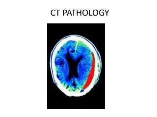
Subdural Hematoma Subdural hematomas are usually the result of a serious head injury. When one occurs in this way, it is called an "acute" subdural hematoma. Acute subdural hematomas are among the deadliest of all head injuries. The bleeding fills the brain area very rapidly, compressing brain tissue. This often results in brain injury.
Stroke Symptoms If you have symptoms of a stroke, seek emergency medical care. General symptoms of a stroke include: Sudden numbness, paralysis, or weakness in your face, arm, or leg, especially on only one side of your body. New problems with walking or balance. Sudden vision changes. Drooling or slurred speech. New problems speaking or understanding simple statements, or feeling confused. A sudden, severe headache that is different from past headaches. Symptoms vary depending on whether the stroke is caused by a clot or bleeding. The location of the blood clot or bleeding and the extent of brain damage can also affect symptoms.
Tumor A brain tumor is a mass or growth of abnormal cells in your brain. Many different types of brain tumors exist. Some brain tumors are noncancerous (benign), and some brain tumors are cancerous (malignant). Brain tumors can begin in your brain (primary brain tumors), or cancer can begin in other parts of your body and spread to your brain (secondary, or metastatic brain tumors).
Multiple Sclerosis • Multiple sclerosis or MS is a disease that affects the brain and spinal cord resulting in loss of muscle control, vision, balance, and sensation (such as numbness). With MS, the nerves of the brain and spinal cord are damaged by one's own immune system. Thus, the condition is called an autoimmune disease.
Trauma • Epidural or extradural hematoma (haematoma) is a type of traumatic brain injury (TBI) in which a buildup of blood occurs between the dura mater (the tough outer membrane of the central nervous system) and the skull. The dura mater also covers the spine, so epidural bleeds may also occur in the spinal column. Often due to trauma, the condition is potentially deadly because the buildup of blood may increase pressure in the intracranial space and compress delicate brain tissue. The condition is present in one to three percent of head injuries.[1] Between 15 and 20% of patients with epidural hematomas die of the injury.[2]
Breast MRI • For a breast MRI, the woman usually lies face down, with her breasts positioned through openings in the table. In order to check breast positioning, the technologist watches the MRI through a window while monitoring for any potential movement. • A breast MRI usually requires the use of a contrast dye that is injected into a vein in the arm before or during the procedure. The dye may help create clearer images that outline abnormalities more easily.
Contrecoup Brain Injury A specific area of brain injury located directly opposite to the site of impact to the head that results from linear violent collisions of the brain with the skull. http://www.neuroskills.com/swfcoup.html
Isodense Subdural Hematoma CT image with contrast demonstrates an isodense subdural hematoma in the left frontoparietal region.
An arteriovenous malformation (AVM) is a congenital defect between the arteries and veins. The condition affects the connection between these blood vessels, and disrupts the flow of blood between them. Although this defect can occur anywhere, AVMs are most common in the brain or spine.
F/X Femoral head MRI
F/X Tarsal Bone MRI