Download
1 / 20
200 likes | 340 Vues
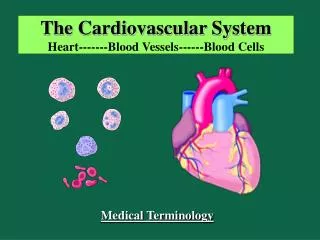
The Cardiovascular System: Blood Vessels Part A. 19. Blood Vessels. Blood is carried in a closed system of vessels that begins and ends at the heart Arteries: carry blood away from the heart Veins: carry blood toward the heart

E N D
The Cardiovascular System: Blood Vessels Part A 19
Blood Vessels • Blood is carried in a closed system of vessels that begins and ends at the heart • Arteries: carry blood away from the heart • Veins: carry blood toward the heart • Capillaries: contact tissue cells and directly serve cellular needs (gas and nutrient)
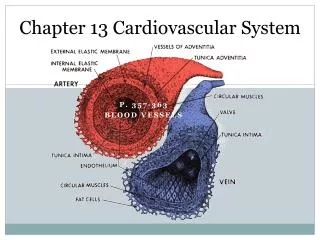
Generalized Structure of Blood Vessels • Arteries and veins are composed of three tunics • tunica interna • tunica media • tunica externa • Lumen – central blood-containing space surrounded by tunics • Capillaries are composed of endothelium with sparse basal lamina
Generalized Structure of Blood Vessels Figure 19.1b
Tunics • Tunica interna (tunica intima) • Endothelial layer that lines the lumen of all vessels • In vessels larger than 1 mm, a subendothelial connective tissue basement membrane is present • Tunica media • Smooth muscle and elastic fiber layer, regulated by sympathetic nervous system
Tunics • Tunica externa (tunica adventitia) • Collagen fibers that protect and reinforce vessels • Larger vessels contain vasa vasorum
Elastic (Conducting) Arteries • Thick-walled arteries near the heart; the aorta and its major branches • Large lumen allow low-resistance conduction of blood • Contain elastin in all three tunics
Muscular (Distributing) Arteries and Arterioles • Muscular arteries – distal to elastic arteries; deliver blood to body organs • Have thick tunica media with more smooth muscle and less elastic tissue • Active in vasoconstriction • Arterioles – smallest arteries; lead to capillary beds
Capillaries • Capillaries are the smallest blood vessels • Walls consisting of a thin tunica interna, one cell thick • Allow only a single RBC to pass at a time • Pericytes on the outer surface stabilize their walls • There are three structural types of capillaries: continuous, fenestrated, and sinusoids
Continuous Capillaries • Continuous capillaries are abundant in the skin and muscles, and have: • Endothelial cells that provide an uninterrupted lining • Adjacent cells that are held together with tight junctions • Intercellular clefts of unjoined membranes that allow the passage of fluids
Continuous Capillaries • Continuous capillaries of the brain: • Have tight junctions completely around the endothelium • Constitute the blood-brain barrier
Continuous Capillaries Figure 19.3a
Fenestrated Capillaries • Found wherever active capillary absorption or filtrate formation occurs (e.g., small intestines, endocrine glands, and kidneys) • Characterized by: • An endothelium riddled with pores (fenestrations) • Greater permeability to solutes and fluids than other capillaries
Fenestrated Capillaries Figure 19.3b
Sinusoids • Highly modified, leaky, fenestrated capillaries with large lumens • Found in the liver, bone marrow, lymphoid tissue, and in some endocrine organs • Allow large molecules (proteins and blood cells) to pass between the blood and surrounding tissues
Sinusoids Figure 19.3c
Capillary Beds • A microcirculation of interwoven networks of capillaries, consisting of: • Vascular shunts – metarteriole–thoroughfare channel connecting an arteriole directly with a postcapillary venule • True capillaries – 10 to 100 per capillary bed, capillaries branch off the metarteriole and return to the thoroughfare channel at the distal end of the bed
Capillary Beds Figure 19.4a
Capillary Beds Figure 19.4b
Vascular Anastomoses • Merging blood vessels, more common in veins than arteries • Arterial anastomoses provide alternate pathways (collateral channels) for blood to reach a given body region • If one branch is blocked, the collateral channel can supply the area with adequate blood supply • Thoroughfare channels are examples of arteriovenous anastomoses