Amino Acids: The Building Blocks of Proteins
Dive into the world of molecules, monomers, and polymers to explore the structure and function of amino acids in proteins. Learn about the unique properties of different amino acids and their role in protein synthesis. Unravel the language of biology through the alphabet of amino acids.

Amino Acids: The Building Blocks of Proteins
E N D
Presentation Transcript
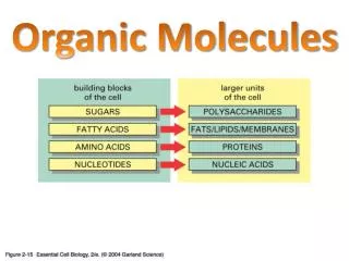
Monomer vs polymer amino acid monomer R is a side group O N-CH-C-OH H 2 R E.g. protein Monomer
O H N-CH-C-OH 2 R R R R R The 20 amino acids found in proteins each have different side chains, R
Each amino acid has at least one carboxyl group O X C OH At cell pH, the carboxyls dissociate to form carboxylate ions O O + + H C X C X O OH
Proteins are the working language of biology The 20 amino acids found in proteins are the alphabet A Let's start with the letter Alanine, Ala, A A All the common amino acids have 3-letter and 1-letter abbreviations
Note asymmetry around the carbon (all 4 side groups are different) Most amino acids have D- and L- isomers* C O O C H H N 3 R *But only L-amino acids are present in typical proteins
Glycine, Gly, G is unusual C O O C H H N 3 H R group is a second H-atom Hence, no asymmetry round carbon No L- and D- isomers
Classes of amino acid R-groups C O O C H H N e.g. Serine, Ser, S 3 CH OH 2 R group Polar, uncharged
Classes of amino acid R-groups C O O C H H N 3 e.g. Aspartic acid, Asp, D CH 2 COO R group* is negatively charged *An additional -COO- group
Classes of amino acid R-groups C O O C H H N 3 e.g. Lysine, Lys, K (CH ) 2 4 NH 3 R group positively charged
R group hydrophobic C O O C O O C H H N C H H N 3 3 CH CH 3 2 e.g. Alanine, Ala, A OH Tyrosine, Tyr, Y
"Special" amino acids C O O C H H N 3 H Glycine, Gly, G (mentioned before) The "baby" (smallest) of the amino acids No special hydrophilic or hydrophobic character
"Special" amino acids C O O C H H N 3 CH 2 SH Cysteine, Cys, C Important in forming disulfide (S-S) crosslinks within a protein
C of Cys DISULFIDE BOND CH C of cysteine 2 S CH H 2 S H S 2H S CH 2 CH 2 of cysteine C of cysteine C
COO +H3N- Special amino acids Proline. This amino acid produces a “kink” in a polypeptide H Proline, Pro, P C CH HN 2 CH H C 2 2 • Pro is an imino acid (not, strictly speaking, • an amino acid
O - + C-O H N 3 O - + H N C-O 3 Forming a peptide bond between 2 amino acids O - C +H3N C C-O + R R 2 1 H20 O C C C N H R R 2 1
+ H N 3 A dipeptide. All peptides and proteins have: and a an amino end carboxyl end O O - C C C-O C N H R R 2 1 C-terminal N-terminal end
Polypeptide structure • Primary • Secondary • Tertiary • Quaternary
1. Primary structure • Amino acid sequence • Disulfide crosslinking within the polypeptide
1. Primary structure: portion of -chain of human hemoglobin N-terminus Glu Lys Val His Leu Thr Pro Glu 3 1 2 5 7 6 4 8 bla bla bla bla bla bla bla bla Tyr Arg bla bla 140 141 C-terminus aa substitution; Val instead of Glu at position 6 is found in the disease sickle cell anemia
2. Secondary stucture of a protein • The folding of portions (domains) of the protein to form: • -helices • -pleated sheets
Secondary structure: -helix Hydrogen bonding 3.6 aa's per turn
Secondary structure: -pleated sheet R R R Edge view R R R R Polypeptide chains H-bond
3. Tertiary structure of protein • Overall folding of: • helices • -pleated sheets and • the regions between them
4. Quaternary structure of protein Ordering of several different polypeptide strands to form a functional protein complex e.g. Hemoglobin