Download
1 / 3
30 likes | 65 Vues
Creative Bioarray cDNA can be used in gene expression and cloning studies, gene mutation analysis, analysis of mRNA alternative splicing and other molecular biology fields.

E N D
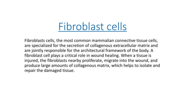
The Principle and Feature of Skin Fibroblast cdna What is Skin Fibroblast cdna? Skin Fibroblast cdna is the main cell component of loose connective tissue, differentiated by mesenchymal cells in the embryonic stage. Fibroblasts are large, clearly defined cells with prominent spindle-shaped or stellate flat structures, with regular oval nuclei and large and prominent nucleoli.According to different functional activity states, the cell can be divided into fibroblasts and fibrocells. fibroblasts have vigorous functional activities, weak basophilic cytoplasm, and obvious protein synthesis and secretion activities.under certain conditions, it can realize mutual transformation with fibroblasts. Fibroblast plays an important role in cell degeneration, necrosis, tissue defect and bone injury repair. In December 2013, researchers at king's college London found different types of skin fibroblast cdna with unique properties that help repair damaged skin and reduce the effects of aging on skin. Features of Skin Fibroblast cdna Skin fibroblasts cdna are the main thins of loose connective tissue. A cell component that is a terminally differentiated cell that produces proteins such as collagen. Fibroblasts have the largest number of large, spindle-shaped or Stella flat cells with many spindles, and their nuclei are regularly ovoid. Its morphology can be changed according to the functional changes of the cell and the physical properties of the attachment site. Skin fibroblasts cdna have large cell bodies, weak basophilic cytoplasm, large oval nuclei, loose chromatin and light staining, and obvious nucleoli.Under electron microscope, abundant rough endoplasmic reticulum, free ribosomes and developed golgi complex were observed in the cytoplasm, indicating that it has the function of protein synthesis and secretion. Skin fibroblasts cdna can synthesize and secrete collagen fiber, elastic fiber, reticular fiber and organic matrix.The procollagen molecules synthesized by it can be polymerized and rearranged by endonuclease to form collagen fibrils with 64nm cycle as the collagen fibrils synthesized and secreted by osteoblasts. The collagen fibrils are bonded to each other to form collagen fibers.Via detecting, synthesis of the two cells secrete collagen fibers are Ⅰ type collagen fiber, exactly the same on the morphological and biochemical structure.Fiber cells in the mature or static state are called fiber cells with small cell body, long spindle shape, underdeveloped rough endoplasmic reticulum and golgi complex.Under the stimulation of trauma and other factors, some fibroblasts can be re-transformed into naive Skin fibroblasts cdna, and their functional activities can be restored to participate in the repair after tissue injury.In addition, there are still a few mesenchymal cells with differentiation potential in the connective tissue, which can proliferate and differentiate into Skin fibroblasts cdna in the case of wound repair.
Principle of Skin Fibroblast cdna Skin fibroblasts cdna take in the required amino acids, such as proline and lysine, and synthesize the prealpha polypeptide chain on the ribosomes of the rough endoplasmic reticulum, which is transported to the golgi complex to form procollagen molecules. Procollagen molecules are carried to the cell surface by secretory vesicles and then released outside the cell by exocytosis. Under the catalysis of procollagen peptidase, the ends of each former alpha polypeptide chain were removed and turned into tropocollagen molecules. Many of the original collagen molecules are arranged in parallel rows and combine to form collagenous fibrils with periodic horizontal stripes. Collagen fibers are formed by combining collagen fibrils with each other. About Creative Bioarray Creative Bioarray is an innovative biotechnology company whose mission focuses on developing unique technologies that provide global scientists with high quality products and satisfactory services to facilitate the investigation of life science researches. We provide a wide range of high quality normal human and animal cells, cell culture medium and reagents, FISH probes, tissue arrays, microorganisms and equipment. In addition, we
also offer series of related services including cell services, biosample services and histology services for the researcher to make their project better and faster.