Understanding Cell Structure and Function: Membranes, Organelles, and Cytoskeleton
170 likes | 292 Vues
This article provides an overview of the structure and function of cell membranes and organelles. It explains the composition of cell membranes, including lipid bilayers and proteins, and the roles of integral and peripheral proteins. Key organelles like mitochondria, ribosomes, the endoplasmic reticulum, Golgi apparatus, lysosomes, and the cytoskeleton are discussed in terms of their functions in cellular maintenance and activity. The fluid mosaic model and the significance of organelles are highlighted to elucidate the dynamic nature of cellular components.

Understanding Cell Structure and Function: Membranes, Organelles, and Cytoskeleton
E N D
Presentation Transcript
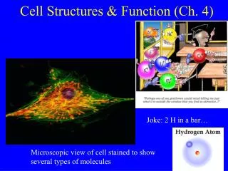
CELL MEMBRANES • The structure of the cell membrane depends on the functions the cell performs. Cell membranes are composed mostly of a lipid bilayer and two type of proteins. Integral proteins are embedded within the membrane. Peripheral proteins are attached to both surfaces of the membrane.
MEMBRANE LIPIDS • One of the major types of lipids in the cell membrane is phos-pholipid. Cells are bathed in an aqueous, or watery, environment. Since the inside of a cell is also an aqueous environment, both sides of the cell membrane are surrounded by water molecules. These water molecules cause the phospholipids of the cell membrane to form two layers- a lipid bilayer.
ORGANELLES • An organelle is a cell component that performs specific functions for the cell. Organelles of a cell maintain the life of the cell. Between the cell membrane and the nucleus lies the cytoplasm, which contains the various organelles of the cell. The organelles are bathed in gelatin-like aqueous fluids called the cytosol. Dissolved in the cytosol are salts, minerals, and organic molecules.
MEMBRANE PROTEINS • Some proteins are attached to the surfaces of the cell membrane. Peripheral proteins are located both in the interior surface and the exterior surface of the cell membrane. Weak bonds link peripheral proteins to membrane lipids or to other proteins that are embedded in the lipid bilayer. The proteins that are embedded in the bilayer are called integral proteins.
Fluid Mosaic Model of Cell Membranes • With the development of new techniques and instruments, including the scanning electron microscope, scientists have discovered that cell membranes are actually very dynamic. These days, scientists use the fluid mosaic model to describe the cell membrane.
Mitochondria • Scattered throughout the cytosolare relatively large organelles called mitochondria. Mitochondria are the sites of chemical reactions that transfer energy from organic compounds to ATP. Mitochondria are usually reactions that occur in a cell. Therefore, mitochondria are usually more numerous in cells that have a high-energy requirement. Mitochondria have their own DNA.
Ribosomes • The most numerous organelles in many cells are the ribsomes. Unlike most other organelles, ribosomes are not surrounded by a membrane. Each ribosome is an assemblage of two organic compounds-proteins and RNA. Inside the cells nucleus, the proteins and RNA re packaged into ribosomes, which are then transported to the cytosol. Ribosomes play important roles in the synthesis ofproteins. Proteins to be used within the cytosol are produced on the ribosomes that are free in the cytosol. Proteins to be inserted into membranes or exported from the cell are produce on the ribosomes that are attached to the endoplasmic reticulum.
Endoplasmic Reticulum • The endoplasmic reticulum, abbreviated ER, is a system of membranes tubules and sacs. The ER functions primarily as an intracellular highway, a path along which molecules move from one part of the cell to another. The amount of ER inside a cell fluctuates, depending on the cells activity. A cell usually contains two types of ER. The two types are Rough endoplasmic reticulum, and Smooth endoplasmic reticulum. Rough ER is prominent in cells that make large amounts of proteins to be exported from the cell or inserted into the cell membrane. Smooth ER is involved in the synthesis of steroids in gland cells, the regulation of calcium levels in muscle cells, and the breakdown of toxic substances by liver cells.
Golgi Apparatus • The Golgi apparatus is the processing, packaging, and secreting of organelle of the cell. Like the endoplasmic reticulum, the Golgi apparatus is a system of membranes.
Lysosomes • Lysosomes are small, spherical organelles that enclose hydrolytic enzymes within single membranes. These enzymes can digest proteins, carbohydrates, lipids, DNA, and RNA. They may also digest old organelles as well as viruses and bacteria that have been ingested by a cell. Lysosomes are common in the cells of animals, fungi, and protists, but they are rare in plant cells. In some multi-cellular organisms, lysosomes play a role during early development. For example, the human hand begins a solid structure in the embryo. As the embryo develops, lysosomal enzymes selectively destroy tissue to from the spaces between the fingers.
Cytoskeleton • Just as your body depends on your skeleton to maintain its shape and size, so a cell needs a structure to maintain its shape and size. In many cells, that structure is the cytoskeleton, a network of long protein strands located in the cytosol. like ribosomes, these strands are not surrounded by membranes. In addition to providing support, the cytoskeleton participates in the movement of organelles within the cytosol. two major component of the cytoskeleton are microfilaments and microtubules. • Microfilaments are threads made of a protein called actin. Each microfilament consists of many actin molecules that are linked together to form a polymer chain. Microfilaments constitute the smallest strands that make up the cytoskeleton. They contribute to cell movement and play a role of in the contraction of muscle cells. • The largest strands of the cytoskeleton are hollow tubes known as microtubules. In many cells, microtubules extend outward from a central point near the nucleus to various sites near the cell membrane. When a cell is about to divide, bundles of microtubules come together and extend across the cell. These bundles, known as spindle fibers, are thick enough to be visible with a light microscope.
Cilia and Flagella • Cilia and Flagella are hair-like organelles that extend from the surface of the cell, where they assist in movement. Because of the variety of roles they play, cilia and flagella can be found in many eukaryotic cells. When these organelles are short and present in large numbers on a cell, they are called cilia. The external surfaces of many unicellular organisms are covered with cilia. The movements of the cilia propel these tiny organisms through the water as they search for food or escape from predators. Cilia are also found on the surfaces of cells in multi-cellular organisms. The cells lining your respiratory tract, for example, bear numerous cilia that trap particles and debris from the air you inhale. As these cilia move, they sweep the trapped materials back up to your throat, where they are removed from your respiratory tract when you swallow.
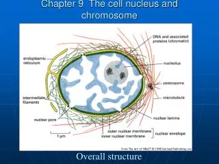
Nucleus • The nucleus is often the most prominent structure within a eukaryotic cell. It maintains its shape with the help of a protein skeleton known as the nuclear matrix. When a cell is about to divide, the chromatin strands coil up and become densely packed, forming chromosomes. The nucleus stores hereditary information in its DNA. The nucleus is also the site where RNA is copied from DNA. In turn, RNA directs the synthesis of proteins, a process that occurs in the cytosol, as you have read. This means that RNA must travel from the nucleus to the cytosol before it can direct protein synthesis. The Nucleus is the site where ribosomes are synthesized and partially assembled before they pass through the nucleuapores to the cytosol.
Cell Wall • Plant cells are covered by a rigid cell wall that lies outside the cell membrane. The rigidity of cell walls helps support and protect the plant. Cell walls contain long chains of cellulose. The cellulose is embedded in proteins and other carbohydrates that harden the entire structure. Pores in the cell wall allow ions and molecules to enter and exit the cell. When the cell reaches its full size, a secondary cell wall may develop.
Vacuoles • Vacuoles are a second common characteristic of plant cells. These fluid-filled organelles store enzymes and metabolic wastes. In fct, some vacuoles may occupy 90 percent of a plant cell’s volume, pushing all the other organelles up against the cell membrane. Some of the wastes stored by vacuoles are toxic and must be kept away from the rest of the cell.
Plastids • A third distinguishing feature of plant cells is the presence of a plastids. Plastids are organelles that, like mitochondria and the nucleus, are surrounded by two membranes and contain DNA. The most familiar type of plastid is the chloroplast. Each chloroplast encloses a system of flattend, membranous sacs called thylakoids. Chloroplasts are organelles in a plant cell in which the energy of sunlight is converted into chemical energy in organic compounds. This conversion occurs in the thylakoids during the process of photosynthesis. Chloroplasts contain large amounts of a green pigment that gives their green color.