Download
1 / 51

E N D
Pathology of Head Injury The goal of mankind is knowledge, which is is inherent in man. No knowledge comes from outside: it is all inside. What man 'learns' is really what he “ discovers ” by taking the cover off his own soul. Swami Vivekananda “ Education” - Eduse (latin) to bring out..
CPC07-4.3.5-CNS-Head Injury <ul><li>Robert is a 62 year old recently retired from QLD railways. </li></ul><ul><li>He lives in Cairns with his wife Rose and their son Aiden who is 40 yrs old with Downs syndrome. </li></ul><ul><li>Presenting symptoms: </li></ul><ul><li>He has fallen from a ladder whilst picking mangoes. </li></ul><ul><li>His wife found him unconscious in the back yard. </li></ul><ul><li>On arrival at the A&E department he is conscious but appears confused . He is complaining of a pain in his L arm. </li></ul> CPC07-4.3.5-CNS-Head Injury <ul><li>Robert is a 62 year old recently retired from QLD railways. </li></ul><ul><li>He lives in Cairns with his wife Rose and their son Aiden who is 40 yrs old with Downs syndrome. </li></ul><ul><li>Presenting symptoms: </li></ul><ul><li>He has fallen from a ladder whilst picking mangoes. </li></ul><ul><li>His wife found him unconscious in the back yard. </li></ul><ul><li>On arrival at the A&E department he is conscious but appears confused . He is complaining of a pain in his L arm. </li></ul>
Examination: <ul><li>General appearance : confused to place and time; no memory of fall or period preceeding fall; drooping R side face and R side of body </li></ul><ul><li>CNS GCS 13 Pupils R>L sluggish response [AVPU]; </li></ul><ul><li>Boggy Haematoma L temporo- parietal area. Gross dysphasia </li></ul><ul><li>Differential Diagnosis </li></ul><ul><ul><li>Head injury with cerebral injury secondary to: </li></ul></ul><ul><ul><ul><li>CVA;embolic </li></ul></ul></ul><ul><ul><ul><li>CVA haemorrhagic </li></ul></ul></ul><ul><ul><ul><li>Trauma to L arm ?# </li></ul></ul></ul><ul><ul><li>Increased intracranial pressure? ? Brain herniation ? </li></ul></ul> Examination: <ul><li>General appearance : confused to place and time; no memory of fall or period preceeding fall; drooping R side face and R side of body </li></ul><ul><li>CNS GCS 13 Pupils R>L sluggish response [AVPU]; </li></ul><ul><li>Boggy Haematoma L temporo- parietal area. Gross dysphasia </li></ul><ul><li>Differential Diagnosis </li></ul><ul><ul><li>Head injury with cerebral injury secondary to: </li></ul></ul><ul><ul><ul><li>CVA;embolic </li></ul></ul></ul><ul><ul><ul><li>CVA haemorrhagic </li></ul></ul></ul><ul><ul><ul><li>Trauma to L arm ?# </li></ul></ul></ul><ul><ul><li>Increased intracranial pressure? ? Brain herniation ? </li></ul></ul>
Control your senses and you are beyond trouble, Let them loose and you are beyond help…! Lao Tzu Control your senses and you are beyond trouble, Let them loose and you are beyond help…! Lao Tzu
Pathology of Head Injury Dr. Shashidhar V.M. Senior Lecturer & Head of pathology Pathology of Head Injury Dr. Shashidhar V.M. Senior Lecturer & Head of pathology
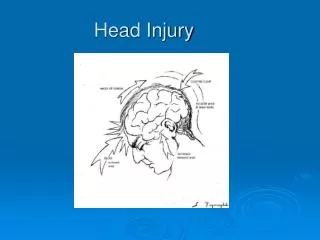
Head Injury Background <ul><li>Skull - Hard casing around soft & delicate Brain Tissue. </li></ul><ul><li>Anterior & posterior compartments separated by tough fibrous coverings </li></ul><ul><li>Rich blood supply </li></ul><ul><li>Neural tracts at base of skull </li></ul> Head Injury Background <ul><li>Skull - Hard casing around soft & delicate Brain Tissue. </li></ul><ul><li>Anterior & posterior compartments separated by tough fibrous coverings </li></ul><ul><li>Rich blood supply </li></ul><ul><li>Neural tracts at base of skull </li></ul>
Head Trauma: Brain injury <ul><li>Two types: Missile & non-missile (blunt) injury </li></ul><ul><ul><li>Common Cause : Traffic accidents & falls </li></ul></ul><ul><li>Primary damage (Coup & Contra-Coup) </li></ul><ul><ul><li>focal lesions (contusions and lacerations) </li></ul></ul><ul><ul><li>diffuse axonal injury </li></ul></ul><ul><li>Secondary damage (after the injury) </li></ul><ul><ul><li>Intracranial haematomas, Oedema </li></ul></ul><ul><ul><li>Intracranial herniation, infarction and infection </li></ul></ul><ul><li>Complications </li></ul><ul><ul><li>epilepsy, post-traumatic dementia </li></ul></ul><ul><ul><li>persistent vegetative state </li></ul></ul> Head Trauma: Brain injury <ul><li>Two types: Missile & non-missile (blunt) injury </li></ul><ul><ul><li>Common Cause : Traffic accidents & falls </li></ul></ul><ul><li>Primary damage (Coup & Contra-Coup) </li></ul><ul><ul><li>focal lesions (contusions and lacerations) </li></ul></ul><ul><ul><li>diffuse axonal injury </li></ul></ul><ul><li>Secondary damage (after the injury) </li></ul><ul><ul><li>Intracranial haematomas, Oedema </li></ul></ul><ul><ul><li>Intracranial herniation, infarction and infection </li></ul></ul><ul><li>Complications </li></ul><ul><ul><li>epilepsy, post-traumatic dementia </li></ul></ul><ul><ul><li>persistent vegetative state </li></ul></ul>
Brain Injury Morphologic Types: <ul><li>Concussion: </li></ul><ul><ul><li>No visible bleed, microscopic diffuse/widespread neuronal stress/damage. </li></ul></ul><ul><li>Contusion: </li></ul><ul><ul><li>Localized, Visible Injury with bleeding (Bruise) </li></ul></ul><ul><li>Laceration: </li></ul><ul><ul><li>Visible linear tear in brain tissue. </li></ul></ul> Brain Injury Morphologic Types: <ul><li>Concussion: </li></ul><ul><ul><li>No visible bleed, microscopic diffuse/widespread neuronal stress/damage. </li></ul></ul><ul><li>Contusion: </li></ul><ul><ul><li>Localized, Visible Injury with bleeding (Bruise) </li></ul></ul><ul><li>Laceration: </li></ul><ul><ul><li>Visible linear tear in brain tissue. </li></ul></ul>
Missile Injury: Types <ul><li>Depressed Injury – skull fracture contusion but no entry to brain. </li></ul><ul><li>Penetrating Injury – enters but not exit. Infection. </li></ul><ul><li>Perforating injury – Missile enters and exits. Large exit wound, extensive haemorrhage, risk of infection & epilepsy. </li></ul> Missile Injury: Types <ul><li>Depressed Injury – skull fracture contusion but no entry to brain. </li></ul><ul><li>Penetrating Injury – enters but not exit. Infection. </li></ul><ul><li>Perforating injury – Missile enters and exits. Large exit wound, extensive haemorrhage, risk of infection & epilepsy. </li></ul>
. Missile Injury: The “blast effect” of a high-velocity projectile causes an immediate increase in supratentorial pressure and results in death because of impaction of the cerebellum and medulla into the foramen magnum. A low-velocity projectile increases the pressure at a more gradual rate through hemorrhage and edema.
. Non-missile / Blunt Head Injury: <ul><li>Primary Injury: </li></ul><ul><ul><li>Coup & Contra-Coup </li></ul></ul><ul><ul><li>Focal damage-concussion, contusion, </li></ul></ul><ul><ul><li>Diffuse axonal injury. </li></ul></ul><ul><li>Secondary Injury: </li></ul><ul><ul><li>Concussion </li></ul></ul><ul><ul><li>Epidural/subdural Hematoma </li></ul></ul><ul><ul><li>Oedema </li></ul></ul><ul><ul><li>Infection </li></ul></ul><ul><li>Post Traumatic Complications: </li></ul><ul><ul><li>Epilepsy </li></ul></ul><ul><ul><li>Dementia </li></ul></ul><ul><ul><li>Vegetative state – Coma. </li></ul></ul>Coup Contre Coup
. Pathogenesis of Injury: <ul><li>Focal damage </li></ul><ul><ul><li>Contusion – Local injury + haemorrhage. </li></ul></ul><ul><ul><li>Healing by gliosis – gross yellow-brown due to hemosidern. </li></ul></ul><ul><li>Diffuse axonal injury </li></ul><ul><ul><li>Shearing of neurons.Two main components exist: </li></ul></ul><ul><ul><li>small haemorrhagic lesions - in the corpus callosum and dorsolateral quadrant of the brainstem; </li></ul></ul><ul><ul><li>diffuse damage to axons – Microscopic - axonal retraction balls & anterograde degeneration. </li></ul></ul>
. Axonal Injury: A, Hypoxic/ischemic injury in cerebral cortex - "red neurons." shrunken cell B, Axonal spheroids at points of axonal disruption C, Swollen cell body and peripheral dispersion of Nissl substance (chromatolysis) H&E Stain.
. Mechanisms of diffuse injury: <ul><li>Excitatory amino acids (EAA) </li></ul><ul><ul><li>Elevation of glutamate and aspartate after Traumatic Brain Injury </li></ul></ul><ul><ul><li>EAAs can cause cell swelling, vacuolization, and neuronal death. Through influx of chloride and sodium, calcium. Decrease ATP or increase free radical production. </li></ul></ul><ul><li>Endogenous opioids & Other alterations: </li></ul><ul><ul><li>behavioral suppression - Activation of muscarinic cholinergic systems in pons </li></ul></ul><ul><ul><li>Decreased glucose utilization further brain injury - increased catecholamines </li></ul></ul><ul><ul><li>Extracellular potassium leading to edema </li></ul></ul><ul><ul><li>Increased cytokines Inflammation </li></ul></ul><ul><ul><li>Decreased intracellular magnesium calcium influx. </li></ul></ul><ul><ul><li>Increased protein kinases astrocyte proliferation </li></ul></ul>
. Head Injury: <ul><li>Multiple contusions involving the inferior surfaces of frontal lobes, anterior temporal lobes, and cerebellum. </li></ul>
. Head Injury: <ul><li>extensive blunt force trauma to the head in a vehicular accident. Mainly the gyri are affected with hemorrhage from contusions and lacerations. </li></ul>
. The greatest religion is to be true to your own nature. Swami Vivekananda
. Intracranial hematoma: Pathological Mechanisms & clinical manifestations No special features Increased intracranial pressure with focal deficits; usually fatal Profound coma , usually rapidly fatal Cortical contusions Rupture of small intrinsic vessels with intracerebral haematoma 'Burst lobe' with intracerebral haematoma contusions and subdural haematoma Cerebral hemisphere Meningeal irritation with a rapid increase in intracranial pressure Arterial rupture Subarachnoid space Acute presentation with a rapid increase in intracranial pressure Chronic presentation with personality change, memory loss and confusion, particularly in the elderly Rupture of venous sinuses or small bridging veins due to torsion forces Subdural space Lucid interval followed by a rapid increase in intracranial pressure Skull fracture with arterial rupture, e.g. middle meningeal artery Epidural /Extradural Clinical manifestations Mechanism Site
. Epidural Hematoma <ul><li>The dura has been reflected back (with a small portion visible at the lower right) to reveal a subdural hematoma. Such a blood clot is usually the result of trauma with tearing of the bridging veins. </li></ul>
. Subdural Hematoma: With head trauma, the dura moves with the skull, and the arachnoid moves with the cerebrum, . As a result, the bridging veins are sheared causing hematoma in the expansile subdural space.
. Subdural Hematoma <ul><li>bilateral chronic subdural hematoma (brown). Since the bleeding is venous, subdurals can form more slowly and insidiously than arterial hemorrhages. Subdurals are most common in the very young and the elderly. </li></ul>
. Subdural Hematoma: edema & Herniation Flattening Subfalcine Herniation Uncal Herniation
. Head Injury:Healing <ul><li>B - Acute contusions are present in both temporal lobes, with areas of hemorrhage and tissue disruption. </li></ul><ul><li>C - Old contusions are present on the inferior frontal surface of this brain, with a brownish yellow color Gliosis+ Hemosiderin </li></ul>
. Head Injury:Healing <ul><li>Old contusions are present on the inferior frontal surface of this brain, with a brownish yellow color </li></ul><ul><li>Gliosis+ Hemosiderin </li></ul>
. You cannot shake hands with a clenched fist. -Indira Gandhi
. You may get delayed to reach your targets. But every step you take towards your target is step towards victory . KARL MARX
. 45y F, Head injury following car accident.Image shows gross specimen brain. ? Diagnosis <ul><li>Subdural Hemorrhage </li></ul><ul><li>Epidural Hemorrhage </li></ul><ul><li>Cerebral Hematoma </li></ul><ul><li>Sub arachnoid Hemorrhage </li></ul><ul><li>Meningeal hemorrhage </li></ul>
. 45y F, Head injury following head on collision, without seat belt.Arrow shows what type of injury? <ul><li>Sub arachnoid Hemorrhage </li></ul><ul><li>Contra coup injury </li></ul><ul><li>Coupe Injury </li></ul><ul><li>Subdural Hematoma </li></ul><ul><li>Meningeal hemorrhage </li></ul>
. 45y F, Head injury following car accident. Image shows MRI Scan.? Diagnosis <ul><li>Subdural Hemorrhage </li></ul><ul><li>Epidural Hemorrhage </li></ul><ul><li>Intracerebral Hematoma </li></ul><ul><li>Sub arachnoid Hemorrhage </li></ul><ul><li>Intra ventricular hemorrhage. </li></ul>
. 45y F, Head injury following car accident. Image shows CT Scan.? Diagnosis <ul><li>Subdural Hemorrhage </li></ul><ul><li>Epidural Hemorrhage </li></ul><ul><li>Intracerebral Hematoma </li></ul><ul><li>Sub arachnoid Hemorrhage </li></ul><ul><li>Intra ventricular hemorrhage. </li></ul>
. 38y Head injury following vehicle accident. Brain gross - What feature is shown by arrow? <ul><li>Subfalcine herniation </li></ul><ul><li>Uncal herniation </li></ul><ul><li>Tonsillar herniation </li></ul><ul><li>Brainstem hemorrhange </li></ul><ul><li>Subdural hemorrhage </li></ul>
. 38y male driver. Died in car crash. Coroners autopsy Image shows Brain gross - What feature is shown by arrow? <ul><li>Old infarct. </li></ul><ul><li>Laceration. </li></ul><ul><li>Recent infarct. </li></ul><ul><li>Hematoma </li></ul><ul><li>Contusion. </li></ul>
. 45y F, Head injury following car accident.Image shows his Brain ? Diagnosis <ul><li>Subfalcine herniation </li></ul><ul><li>Brain stem hemorrhage </li></ul><ul><li>Uncal herniation </li></ul><ul><li>Cerebral edema </li></ul><ul><li>2, 3 & 4 are correct. </li></ul>2 3 4
. Do not let what you ‘cannot’ do interfere with what you ‘can’ do . — John Wooden
. Sample SAQ: <ul><li>List effects of increased intracranial pressure produced by supratentorial space occupying lesions. </li></ul><ul><li>List the consequences of uncal herniation. </li></ul><ul><li>Describe the effects of downward displacement of the brainstem (central herniation). </li></ul><ul><li>Describe the pathogenesis and consequences of cerebellar tonsil herniation. </li></ul><ul><li>List the types of cerebral edema and discuss their pathogenesis and consequences. </li></ul>
. Head Injury: Clinical severity <ul><li>First Degree </li></ul><ul><ul><li>No Concussion - Bell rung resolution of symptoms within Lucid Interval. </li></ul></ul><ul><li>Second Degree </li></ul><ul><ul><li>Loss of Consciousness <15 sec . </li></ul></ul><ul><ul><li>No resolution within lucid interval </li></ul></ul><ul><li>Third Degree </li></ul><ul><ul><li>Loss of Consciousness >15 Sec. </li></ul></ul><ul><ul><li>increasing severity within lucid interval </li></ul></ul>
. Decisions on Head Injuries <ul><li>MUST SEND TO EMERGENCY IF: </li></ul><ul><ul><li>Blood out of ears &/or nose (from blow to head) </li></ul></ul><ul><ul><li>Any unconscious episode </li></ul></ul><ul><ul><li>Increase in Symptoms : during 20 minute waiting period or when activity resumes </li></ul></ul><ul><li>DO NOT RETURN TO ACTIVITY IF: </li></ul><ul><ul><li>Symptoms are not resolved </li></ul></ul><ul><ul><li>Activity exacerbates headache </li></ul></ul>
. Post-Concussive Syndrome <ul><li>Residual symptoms post concussion including </li></ul><ul><ul><li>Nausea </li></ul></ul><ul><ul><li>Headache </li></ul></ul><ul><ul><li>Tinnitus </li></ul></ul><ul><ul><li>etc. </li></ul></ul><ul><li>These may occur with movement, daily activities, rising from bed etc. </li></ul><ul><li>Must not participate during this period of time </li></ul>
. Second Impact Syndrome <ul><li>Injury while still recovering from initial concussion </li></ul><ul><li>100% morbidity </li></ul><ul><li>Very important that post-concussive syndrome has resolved </li></ul>
. Athlete Return to Activity <ul><li>First Concussion 1º </li></ul><ul><ul><li>1 week post resolution of Symptoms </li></ul></ul><ul><li>Second Concussion or 2 º </li></ul><ul><ul><li>1 month post resolution of symptoms </li></ul></ul><ul><li>Third Concussion or 3 º </li></ul><ul><ul><li>End of Season needs discussion of career </li></ul></ul><ul><li>Start with bike activity - if no symptoms increase functional activity before full return </li></ul><ul><li>Above subject to Dr Discretion </li></ul>
. Religion is the symptom of craving to know Divinity already inside man. Swami Vivekananda