Pathology Review-Term3
1.05k likes | 1.16k Vues
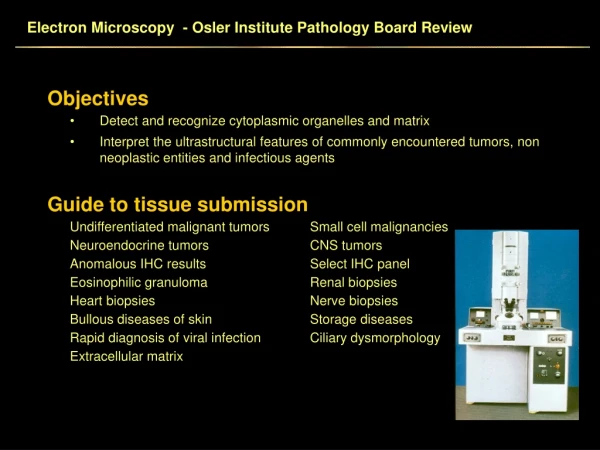
medical school pathology lectures. Year end review of Term3. Endocrine & CNS. Note: Video of this lecture is on on youtube.

Pathology Review-Term3
E N D
Presentation Transcript
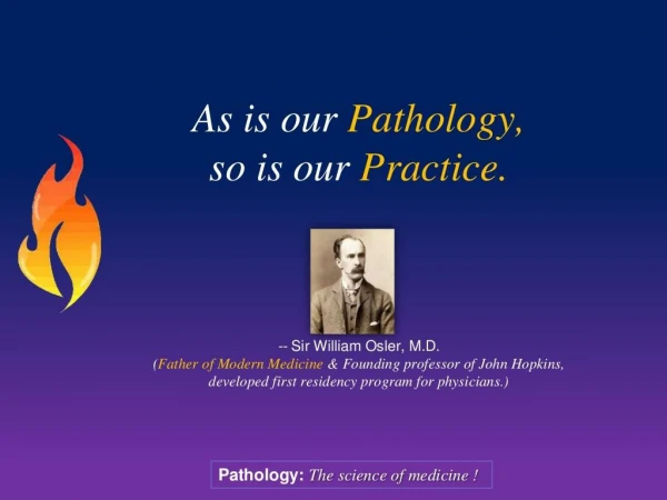
Pathology Review-Term3 As is our Pathology, so is our Practice. -- Sir William Osler, M.D. (Father of Modern Medicine & Founding professor of John Hopkins, developed first residency program for physicians.) Pathology: The science of medicine !
CPC31: Week Overview 2013 Term 3 CPC 1 Title: Endocrine 1/1 Thyroid System: Endocrine Aim: To educate students in Clinical, pathology & population study of patients with Endocrine disorders (using example of thyroid disorder). Learning Outcomes: 1. Demonstrate an ability to take a focused History & Students will be able carry out a focused clinical examination of patients to with neck swelling and systemic symptoms 2. Describe the Pathophysiology of common endocrine disorders. 3. Describe the pathophysiology of thyroid disorders. 4. Outline the basic sciences relating to hypothalamic/pituitary/thyroid axis; thyroid function testing; role of anti thyroid antibodies in differentiating thyroid disease 5. Outline first line treatment of thyroid disorders. CPC31: Week Overview 2013 Term 3 CPC 1 Title: Endocrine 1/1 Thyroid System: Endocrine Aim: To educate students in Clinical, pathology & population study of patients with Endocrine disorders (using example of thyroid disorder). Learning Outcomes: 1. Demonstrate an ability to take a focused History & Students will be able carry out a focused clinical examination of patients to with neck swelling and systemic symptoms 2. Describe the Pathophysiology of common endocrine disorders. 3. Describe the pathophysiology of thyroid disorders. 4. Outline the basic sciences relating to hypothalamic/pituitary/thyroid axis; thyroid function testing; role of anti thyroid antibodies in differentiating thyroid disease 5. Outline first line treatment of thyroid disorders.
Head injury, infections, Inflammations & Tumours Central Diabetes Insipidus, Hypogonadism Adenoma, Ischemia (Sheehans), Diabetes insipidus, Atrophy / hyperfunction Hyper/Hypo parathyroidism Graves, Hashimoto, MNG, Cancer. Hypothal: Tertiary Pituitary: Secondary Gland: Primary Cushings, Addisons, Pheochromocytoma Adenoma, carcinoma Tumours, Cysts, DIABETES Adenoma, carcinoma Tumours, Cysts, Atrophy, Dysfunction Tumours, Cysts, Atrophy, Dysfunction Head injury, infections, Inflammations & Tumours Central Diabetes Insipidus, Hypogonadism Adenoma, Ischemia (Sheehans), Diabetes insipidus, Atrophy / hyperfunction Hyper/Hypo parathyroidism Graves, Hashimoto, MNG, Cancer. Hypothal: Tertiary Pituitary: Secondary Gland: Primary Cushings, Addisons, Pheochromocytoma Adenoma, carcinoma Tumours, Cysts, DIABETES Adenoma, carcinoma Tumours, Cysts, Atrophy, Dysfunction Tumours, Cysts, Atrophy, Dysfunction
Hyper – Thyroidism - Hypo • Hyper-Metabolic… • Hypo-Metabolic… Hyper – Thyroidism - Hypo • Hyper-Metabolic… • Hypo-Metabolic…
Nontoxic-Multi Nodular Goitre. A: conspicuous neck mass. B: Coronal section showing numerous irregular nodules, some with hemorrhage. C: Microscopy: variation in the size of the follicles. Note: TSH, T3,T4 Normal (Euthyroid) Nontoxic-Multi Nodular Goitre. A: conspicuous neck mass. B: Coronal section showing numerous irregular nodules, some with hemorrhage. C: Microscopy: variation in the size of the follicles. Note: TSH, T3,T4 Normal (Euthyroid)
Carcinoma of Thyroid Type (%) age spread Prognosis Papillary 60-70 young adults 20-40 (<45y) Lymphatic, to local nodes Follicular 20-25 Young-middle 4050 (>45) Blood stream, Good with radioespecially to bone iodine therapy. Papillary Carcinoma: Psammoma Body Excellent Carcinoma of Thyroid Type (%) age spread Prognosis Papillary 60-70 young adults 20-40 (<45y) Lymphatic, to local nodes Follicular 20-25 Young-middle 4050 (>45) Blood stream, Good with radioespecially to bone iodine therapy. Papillary Carcinoma: Psammoma Body Excellent
SIADH: Sy of Inappropriate ADH secretion SIADH • • • • • Too Much ADH Water Intoxication Low Serum Sodium Low Serum Osmolality High Urine Osmolality Diabetes Insipitus • • • • • Too Little ADH Dehydration High Serum Sodium High Serum Osmolality Low Urine Osmolality SIADH: Sy of Inappropriate ADH secretion SIADH • • • • • Too Much ADH Water Intoxication Low Serum Sodium Low Serum Osmolality High Urine Osmolality Diabetes Insipitus • • • • • Too Little ADH Dehydration High Serum Sodium High Serum Osmolality Low Urine Osmolality
Adrenal Glands: • Cortex (glands) – Glomerulosa - Mineralocorticoids – Fasciculata - Glucocorticoids – Reticularis – Gonadal hormones • Medulla (Neural) – Chromaffin cells & sympathetic nerve endings - Noradrenaline Adrenaline (epinephrine) • Pathology: common. – – – – – Pheochromocytoma – medulla hypertension. Addison‟s disease* Cushing‟s syndrome* & Disease*. Conn‟s syndrome*. Congenital adrenal hyperplasia(21-hydroxylase deficiency) Precocious puberty Excess Androgens Adrenal Glands: • Cortex (glands) – Glomerulosa - Mineralocorticoids – Fasciculata - Glucocorticoids – Reticularis – Gonadal hormones • Medulla (Neural) – Chromaffin cells & sympathetic nerve endings - Noradrenaline Adrenaline (epinephrine) • Pathology: common. – – – – – Pheochromocytoma – medulla hypertension. Addison‟s disease* Cushing‟s syndrome* & Disease*. Conn‟s syndrome*. Congenital adrenal hyperplasia(21-hydroxylase deficiency) Precocious puberty Excess Androgens
Pheochromocytoma: • • • • • • Tumor of medullary Chromaffin cells. Increased catecholamines Secondary hypertension. Young age. May be familial (MEN syndrome). Increased Urinary VMA Pheochromocytoma: • • • • • • Tumor of medullary Chromaffin cells. Increased catecholamines Secondary hypertension. Young age. May be familial (MEN syndrome). Increased Urinary VMA
. Waterhouse-Friderichsen Sy • • • • • • Acute hemorrhagic necrosis (apoplexy). Shock/Septicemia Lack of aldosterone Salt & water loss K Hypovolemic shock Hypoglycemia. K The adrenals from a child dying of meningococcal septicaemia are destroyed by haemorrhage.
. Addison’s Disease: • • • • • • • • Chronic adrenal insufficiency. anorexia, weight loss, vomiting Weakness, lethargy, hypotension skin pigmentation hyponatraemia with hyperkalaemia chronic dehydration, sexual dysfunction. Low plasma cortisol. ACTH high (primary) or low (sec) • Autoimmune 70%, • Infections (TB, fungal), tumours,
. Education is what remains after we have forgotten all the facts taught in the class! --
. CPC32: Week Overview 2013 Term 3 CPC 2 Endocrine 2 - Diabetes System: Endocrine Aim: To educate students in: Clinical, Pathology & population study of patients with Diabetes Mellitus Learning 1. Demonstrate an ability to carry out a focused History & Outcomes clinical examination of patients with diabetes. Students will be 2. Describe the pathophysiology of Diabetes Mellitus able to Types 1 + 2 and their complications. 3. Outline the Basic sciences relating to endocrine function. 4. Explain the abnormalities of Blood supply common in diabetic patients including - ischaemia, infarction & necrosis. 5. Outline the epidemiology and aetiology of Diabetes Mellitus in Australia and world wide. 6. Outline first-line management and demonstrate an understanding of the process of care of a diabetic patient.
. Most likely .. What type of DM ? II NIDDM II GDM I IDDM Sec IDDM Sec IDDM I LADA Sec IDDM I IDDM LADA MODY 1. 56 year male obese 2. 30 year female following pregnancy 3. 8 year old boy, poor growth, DKA. 4. 24 year female Cushing‟s sy 5. 68 Year male following Ca. pancreas. 6. 32 male, DM, BMI 18, Anti-GAD +ve. 7. 34 year male, extensive tuberculosis. 8. 12 year old female following viral fever 9. 41y DM2, BMI 17.1, HbA1c 14.1, DKA 10.15y male, BMI 16.2, recurrent infect.
. DM Questions • • • • • • • Definition? types common? Diagnosis? Primary & Sec? Congenital? Gestational? Monogenic? MODY, LADA, drugs? List functions of Insulin? Antagonists? Etiology & Pathogenesis of Type 1 & 2. Stages of DM & their pathological basis? Obesity & Insulin resistance * – FFAs, PKC, Adipkines, PPARγ – Inflammation & Insulin resistance. • Mechanism of β cell destruction type 1, 2. • Islet Amyloid PolyPeptide (IAPP)?
. DM Questions • MODY & LADA – pathogenesis, subtypes. • Pathogenesis of Complications: – – – – – – Mechanism: AGEs, Activate of PKC, & Polyols. Infections – common & pathogenesis. Foot ulcer, Retinopathy (prol & non-prol), Neuropathy? Central, peripheral, autonomic… Difference Angiopathy Micro & Macro? MI, Stroke. Diabetic Nephropathy – albuminuria, KW lesion, Papillary Necrosis, Pyelonephritis, CRF. – Hypertension, Cataract, • Metabolic: Diabetic Coma, DKA, HONK **
. Insulin Anabolic Steroid GLUT4 * only these tissues….!
. Obesity & Insulin resistance. Peripheral Res. Diabetes is a state of inflammation
. GIP : glucose-dependent insulinotropic polypeptide (GIP) GLP-1: glucagon-like peptide-1 (GLP-1) DPP 4: enzyme dipeptidyl peptidase-4 (DPP-4) – breaks down GIP & GLP-1
. Diabetes Classification: (not a single disease) – Type I – IDDM / Juvenile – 5-10%. – Type II – NIDDM /Adult onset – 90-95%. – MODY – 5% Maturity Onset Diabetes of Youth • Genetic, sub types MODY 1–6 (3 & 2 common), – LADA – Latent Autoimmune Diabetes in Adults (LADA) – Gestational Diabetes Mellitus. – Other. (neonatal diabetes, – Insulin gene defects) Endocrinopathy, Downs Sy. – Excess hyperglycemic stimulus (drugs & disease). • Cushings, Phaeochromocytoma, acromegaly, Steroid therapy. – Beta cell destruction: • Pancreatitis/tumors/Hemochromatosis – Bronze diabetes. • Infectious – congenital rubella, CMV, TB,
. Pathogenesis of Type I DM Insulin deficiency ß cell Destruction Antibodies: Islet cell Ab - ICA Insulin Auto Ab - IAA Glut. Acid Decarb - GAD65 Autoimmune Insulitis Ab to ß cells/insulin Environment Viral infe..? Genetic HLA-DR3/4 Secondary DM Inflammation, Tumor, Infection Trauma Pancreatitis
. ? β cell dysfunction Relative Insulin Def. Type II Pathogenesis IDDM Β cell Exhaustion
. Type-I Insulitis: Lymphocytic infiltrate within islets. Type-II Islet Hyalinization: Central hyaline deposits replacing dead beta cells (only in late stage…!)
. DM Microangiopathy – pathogenesis Normal Glucose Glycosylation BM damage leak „AGE‟ deposition PATHOGENESIS OF DM COMPLICATIONS: 1. Chronic Hyperglycemia. 2. Glycosylation of BV B. membrane 3. Leakage of proteins, excess BM matrix. 4. Narrow, Diabetic thick, fragile, Leaky BV + Inflam. 5. Leakage of LDL, protein, angiogenesis. • Ischemia • Proteinuria (kidney) • Micro Aneurysms (retina) • Atherosclerosis.
. “A man must be big enough to admit his mistakes, smart enough to profit from them, and strong enough to correct them!” --John C. Maxwell
. CPC33: Week over veiw 2013 Term 3 CPC 3 Title: Neurology 1 – Head injury, Stroke. System: CNS-Neurology Aim: To educate students in: Clinical, Pathology & population study of patients with Head Injury, including non traumatic brain injury & stroke. Learning 1. Demonstrate an ability to take a focused history & perform a relevant Outcomes: and focused clinical examination of patients with loss of Students will be consciousness/alteration in neurological function able to 2. Describe the Pathophysiology of cerebrovascular accidents. 3. State the Pathophysiology of hypertension (review). 4. Recall the components of Basic sciences relating to function of the brain. 5. Define the Epidemiology and Pathology of cerebrovascular disease (stroke). 6. Describe the Epidemiology of neurological diseases in Australia and world wide. 7. Define the first line management of patients with impaired Glasgow Coma Scores 8. Demonstrate an understanding of the complications of intra-cerebral events
. Learning Objectives: • CNS: Functional Anatomy, Physiology & Blood supply – Revision • Stroke: Definition, types, etiology, clinical features, complications. • Brain Ischemia: TIA, Embolic, Hemorrhagic & Ischemic. • Healing in brain tissue – liquifactive necrosis, gliosis. • • • • Head Injury: types, pathophysiology, clinical & complications. Concussion, Contusion, Laceration. Hemorrhage: Epidural, Subdural, Subarachnoid, intra cerebral. Brain herniation – types & clinical features. Subfalcine, uncal, tonsillar. • Hypertension: Pathophysiology, diagnosis & Complications *Self Study • Hypertension & CNS damage, Hypertensive encephalopathy. • Epilepsy: Overview, types, pathophysiology, Clinical *Self Study
. Blunt Head Injury: • Primary Injury: – Coup & Contra-Coup – Focal damage-concussion, contusion, – Diffuse axonal injury. • Secondary Injury: – – – – Concussion Epidural/subdural Hematoma Oedema Infection • Post Traumatic Complications: – Epilepsy – Dementia – Coma. Contre Coup
. Epidural - Hematoma - Subdural Old age, fall, delayed symptoms Arachnoid Pia fixed to brain Sub arachnoid Hptn, Atherosclerosis
. Subarachnoid Hem: alone is not trauma..! Think of: • Hypertension, • Berry aneurysm, • Atherosclerosis.
. Hypertensive Intracerebral Hem: Sites 1. Putamen-Claustrum 2. Cerebral white matter 3. Thalamus 4. Pons 5. Cerebellum 55% - MCA deep br. 15 10 10 10 Aetiology: Atherosclerosis – most common. Hypertension, smoking, diabetes. Heart disease – Atrial fibrillation Other: Trauma – fat embolism Tumor, Infection Caissons disease – Bends *Pacific.
. Left (Dominant) Hemisphere Stroke: Clinical • • • • • • • Aphasia Right hemiparesis Right-sided sensory loss Right visual field defect Poor right conjugate gaze Dysarthria Difficulty reading, writing, or calculating Diagnosis: Recent cerebral infarction in left MCA distribution. Left cerebral hemisphere shows swelling with compression of the lateral ventricle mainly in the frontal area, due to recent infarct in the Middle Cerebral Artery (MCA) distribution. The brain in the MCA area shows discoloration of the cortex and also blurring between the cortex and white matter.
. ACA stroke. • Paralysis of contralateral foot and leg • Sensory loss over toes, foot and leg • Impairment of gait and stance • Abulia (slowness and prolonged delays to perform acts) • Flat affect, lack of spontaneity, slowness, distractibility • Cognitive impairment, such as perseveration and amnesia • Urinary incontinence Wikipedia: GNU Free Documentation license
. PCA stroke. Peripheral (cortical) • Homonymous hemianopia • Memory deficits • Perseveration (repeat response) • Several visual deficits (cortical blindness, lack of depth perception, hallucinations) Central (penetrating) • Thalamus - contralateral sensory loss, spontaneous pain, mild hemi • Cerebral peduncle - CN III palsy with contralateral hemiplegia • Brain stem - CN palsies, nystagmus, pupillary abnormalities Wikipedia: GNU Free Documentation license
. Infarct Pathogenesis: Hours 1-day 3-day 1 wk. >4wk • • • • • • • • • hypoxia/anoxia. Na/K pump block. Calcium influx. Red neuron, vacuolation. Cell death, karyorrhexis. Inflam- neutrophils, hem. Lympho, Macrophages. Liquefaction cavity Peripheral Gliosis (astrocytes)
. CNS AV Malformations: • Many types: – – – – AV Malformation * Cavernous angioma Telangiectasia Venous angioma • Cause of Seizure disorders & hemorrhage. • Most common congenital vascular malformation. • Typically located in the outer cerebral cortex underlying white matter.
. Hypertension Stroke: Hemorrhagic stroke (new) & Lacunar infarct (old)
. Our greatest glory is not in never falling, but in rising every time we fall. -- Confucius
. CPC34: Week over view 2013 Term 3 CPC Title: Neurology 2 4 System: Neurology Aim: To educate students in: Clinical, Pathology & population study of patients with dementia Learning 1. Demonstrate an ability to take a relevant and focused history and Outcomes: carry out a relevant clinical examination of patients with dementia. (Students will be 2. Carry out a competent physical examination of neurological system. able to) 3. Describe the Pathophysiology & Pathology of common causes of dementia (Alzheimer’s Disease, multi infarct dementia) 4. Recognsise rare neurological causes of dementia (Huntington’s disease, CJD, HIV). 5. Outline the basic sciences relating to structure and function of the brain. 6. Describe the difference between age associated memory impairment, mild cognitive impairment and Alzheimer’s disease 7. Outline the epidemiology of Alzheimer’s disease and other causes of dementia in Australia. 8. Explain the first line management of common causes of dementia 9. Outline ethical dilemmas in caring for patients with dementia
. Broca‟s area - Cingulate and Parahippocampal gyri. Hippocampus: where short-term memories are converted to longterm memories Thalamus: receives sensory and limbic information and sends to cerebral cortex (cognition) Limbic system: controls emotions and instinctive behavior (includes the hippocampus and parts of the cortex)