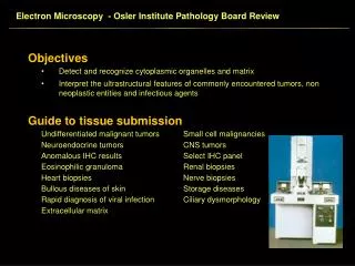
Electron Microscopy - Osler Institute Pathology Board Review
Electron Microscopy - Osler Institute Pathology Board Review. Primer of Organelles Cytoplasm Annulate lamellae Cellular immaturity. Electron Microscopy - Osler Institute Pathology Board Review. Primer of Organelles Cytoplasm Cylindric confronting cisternae

Electron Microscopy - Osler Institute Pathology Board Review
E N D
Presentation Transcript
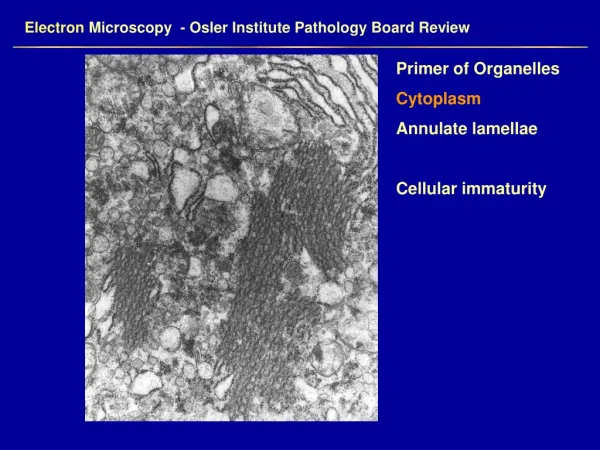
Electron Microscopy - Osler Institute Pathology Board Review Primer of Organelles Cytoplasm Annulate lamellae Cellular immaturity
Electron Microscopy - Osler Institute Pathology Board Review Primer of Organelles Cytoplasm Cylindric confronting cisternae Ring shaped and tubular forms in HIV infection
Electron Microscopy - Osler Institute Pathology Board Review • Primer of Organelles • Cytoplasm • Microtubules • Cell processes of neuroblastoma • Intracisternal microtubules • Melanoma • Extraskeletal chondrosarcoma
Electron Microscopy - Osler Institute Pathology Board Review Primer of Organelles Cytoplasm Myosin and Actin Myosin - 15 nm Actin – 6 -8 nm
Electron Microscopy - Osler Institute Pathology Board Review Primer of Organelles Cytoplasm Cytokeratin filaments Squamous cell carcinomas
Electron Microscopy - Osler Institute Pathology Board Review Primer of Organelles Cytoplasm Vimentin filaments Mesenchymal cells, endothelial cells, tumors Abundant in rhabdoid tumors of the kidney Carcinoid tumor with intracytoplasmic vimentin filaments
Electron Microscopy - Osler Institute Pathology Board Review Primer of Organelles Cytoplasm Intermediate filaments Aggregates of smooth muscle actin Infantile digital fibromatosis
Electron Microscopy - Osler Institute Pathology Board Review Primer of Organelles Cytoplasm Glial filaments 51 – 52 KD glial fibrilllary acidic protein Astrocytic tumors Plaques of multiple sclerosis Rosenthal fibers – pilocytic astrocytoma
Electron Microscopy - Osler Institute Pathology Board Review • Primer of Organelles • Endoplasmic reticulum • Rough endoplasmic reticulum • Abundant RER in: • Plasma cells • Acinar cell neoplasms
Electron Microscopy - Osler Institute Pathology Board Review Primer of Organelles Cytoplasm Smooth Endoplasmic reticulum Ovarian sex cord tumors Lipid cell tumors Hilus cell tumor of ovary Luteomas Sertoli adenoma of testis Adrenal cortical tumors Sebaceous gland neoplasms
Electron Microscopy - Osler Institute Pathology Board Review Primer of Organelles Cytoplasm Intercellular junctions Junctional complexes – cytoplasmic lumina Desmosomes - carcinomas Primitive junctions – sarcomas, carcinomas
Electron Microscopy - Osler Institute Pathology Board Review Primer of Organelles Cytoplasm Lamellar cytoplasmic inclusions Myelinosomes Bronchioloalveolar cell carcinoma Type II pneumocyte
Electron Microscopy - Osler Institute Pathology Board Review Primer of Organelles Cytoplasm Langerhan’s cell (Birbeck) granules Eosinophilic granuloma
Electron Microscopy - Osler Institute Pathology Board Review Primer of Organelles Cytoplasm Lipid Hibernomas Steroid secreting cells Clear cell carcinoma of kidney Malignant fibrous histiocytoma
Electron Microscopy - Osler Institute Pathology Board Review Primer of Organelles Cytoplasm Lysosomes Secondary lysosomes Granular cell tumors Myelomonocytic leukemia Malignant histiocytosis