Download

1 / 15
170 likes | 445 Vues
Osler Institute – Pathology Board Review: Electron Microscopy. EM of Tumors and Tumor-like Entities. Electron Microscopy - Osler Institute Pathology Board Review. Neoplasms Oncocytoma Salivary gland Parotid Kidney. Electron Microscopy - Osler Institute Pathology Board Review. Neoplasms

E N D
Osler Institute – Pathology Board Review: Electron Microscopy EM of Tumors and Tumor-like Entities
Electron Microscopy - Osler Institute Pathology Board Review • Neoplasms • Oncocytoma • Salivary gland • Parotid • Kidney
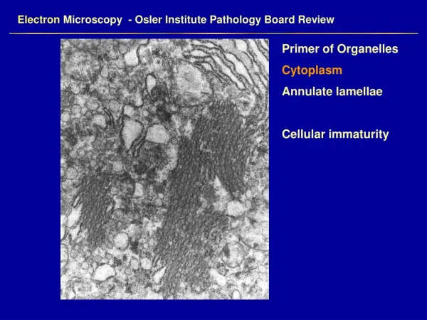
Electron Microscopy - Osler Institute Pathology Board Review • Neoplasms • Oncocytoma • Pleomorphic mitochondria • Lamelliform (plate like) mitochondrial cristae • Dense bodies in mitochondria
Electron Microscopy - Osler Institute Pathology Board Review • Neoplasms • Plasmacytoma • Nuclei • Marginated heterochromatin • Prominent profiles of rough endoplasmic reticulum • Immunoglobulin inclusions in RER and nucleus
Electron Microscopy - Osler Institute Pathology Board Review • Neoplasms • Renal Cell Carcinoma • (conventional type) • Long microvilli • Glycogen • Lipid droplets
Electron Microscopy - Osler Institute Pathology Board Review • Neoplasms • Renal Cell Carcinoma • Chromophobe Type • Microvesicles • Hale’s colloidal iron positive • Papillary (chromophil) Type • Abundant mitochondria
Electron Microscopy - Osler Institute Pathology Board Review • Neoplasms • Seminoma • Wandering, ropy, nucleolus • Finely dispersed heterochromatin • Inconspicuous organelles • Intercellular junctions
Electron Microscopy - Osler Institute Pathology Board Review • Neoplasms • Squamous cell carcinoma • Desmosomes • Tonofilament bundles
Electron Microscopy - Osler Institute Pathology Board Review Small Round Cell Tumors
Electron Microscopy - Osler Institute Pathology Board Review • Neoplasms • Small Round Cell Tumors • Ewings Sarcoma • Dispersed heterochromatin • Small nucleolus
Electron Microscopy - Osler Institute Pathology Board Review • Neoplasms • Small Round Cell Tumors • Ewings Sarcoma • Large pools of glycogen
Electron Microscopy - Osler Institute Pathology Board Review • Neoplasms • Small Round Cell Tumors • Rhabdomyosarcoma • Actin-myosin filaments with Z-band • Primitive sarcomeres
Electron Microscopy - Osler Institute Pathology Board Review • Neoplasms • Small Round Cell Tumors • Rhabdomyosarcoma • Ribosome-filament complexes • Small primitive junctions • External lamina
Electron Microscopy - Osler Institute Pathology Board Review • Neoplasms • Small Round Cell Tumors • Neuroblastoma • Cell processes • Secretory granules • Microtubules • Neurofilaments
Electron Microscopy - Osler Institute Pathology Board Review • Neoplasms • Small Round Cell Tumors • Lymphoma (Follicular Center Cell) • No cell junctions