Download
1 / 4
40 likes | 466 Vues
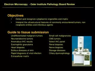
Electron Microscopy - Osler Institute Pathology Board Review. Objectives Detect and recognize cytoplasmic organelles and matrix Interpret the ultrastructural features of commonly encountered tumors, non neoplastic entities and infectious agents Guide to tissue submission

E N D
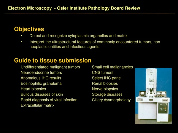
Electron Microscopy - Osler Institute Pathology Board Review • Objectives • Detect and recognize cytoplasmic organelles and matrix • Interpret the ultrastructural features of commonly encountered tumors, non neoplastic entities and infectious agents • Guide to tissue submission • Undifferentiated malignant tumors Small cell malignancies • Neuroendocrine tumors CNS tumors • Anomalous IHC results Select IHC panel • Eosinophilic granuloma Renal biopsies • Heart biopsies Nerve biopsies • Bullous diseases of skin Storage diseases • Rapid diagnosis of viral infection Ciliary dysmorphology • Extracellular matrix
Electron Microscopy - Osler Institute Pathology Board Review • Processing tissue for electron microscopy • 1 mm cubed tissue pieces • Dehydrate, infiltrate, osmicate, embed in plastic • Toluidine blue thick sections • Thin sections – 100 nm on copper grids (silver sections) • Interpretation of electron micrographs • Determine approximate magnification • Identify organelles • Integrate ultrastructural features with clinical history, light microscopy, IHC and molecular diagnostics • Limitations of electron microscopy • Sample size • Multidirectional differentiation • No diagnostic organelles
Electron Microscopy - Osler Institute Pathology Board Review • Primer of Organelles • Nucleus • Abundant heterochromatin – Ewings sarcoma, B cell lymphoma • Cerebriform nuclear contours • Nuclear pseudoinclusions – nuclear pockets • Intranuclear tubular aggregates • Nucleolus • Meandering reticular nucleoli – seminoma, dysgerminoma • Marginated Nucleolus • Increased nucleoli Slide 3
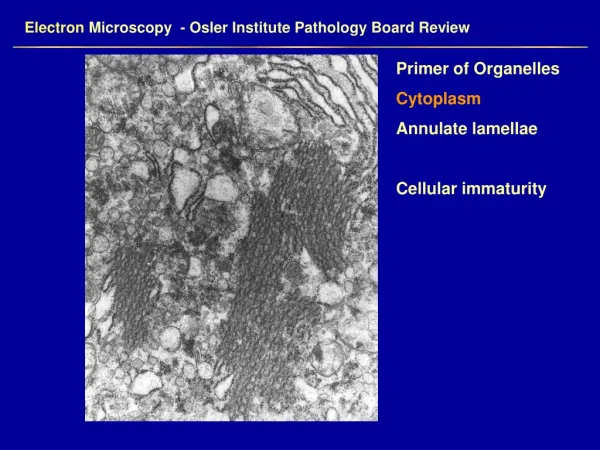
Electron Microscopy - Osler Institute Pathology Board Review • Primer of Organelles • Cytoplasm • Annulate lamellae Cylindrical confronting cisternae • Cytoskeleton Endoplasmic reticulum • Golgi Intercellular junctions • Lamellar cytoplasmic inclusions Lipid • Langerhan’s cell granules Lysosomes • Melanosomes Microvilli • Mitochondria Ribosomes and polyribosomes • Secretory granules Tubular reticular structures • Weibel Palade bodies • Extracellular Matrix • Amyloid • Collagen