Cell Structure and function
E N D
Presentation Transcript
Cell Structure and function Chapter 4
Schedule and Announcements • Quiz Friday over organelles and cell structure • Lab Thursday over cell structure and function • Test 3- Tuesday 10/13 over Organelles and transport • Test 4- Friday 10/16 over Q1 material
Student Learning Goals - BiologyCell TheorySC.912.L.14.1 Goal:Describe the scientific theory of cells and relate the history of its discovery to the process of science. 4 – Research and investigate the scientific theory of cells and relate the history of its discovery to the process of science. 3 - Describe the scientific theory of cells and relate the history of its discovery to the process of science. 2 – Define the scientific theory of cells and relate the history of its discovery to the process of science. 1 – Recognize the scientific theory of cells and relate the history of its discovery to the process of science.
Learning objectives • Explain why the cell is considered to be the basic unit of life. • Name the scientists who first observed living and nonliving cells. • State the principles of the cell theory. • Summarize the research that led to the development of the cell theory. • Explain the relationship between cell shape and cell function. • Identify the factor that limits cell size. • Describe the 3 basic parts of a cell
Learning objectives 8. Compare and contrast prokaryotic and eukaryotic cells. 9. Analyze the relationship among cells, tissues, organs, organ systems, and organisms 10. Describe the structure and function of a cell’s plasma membrane 11. Summarize the role of the nucleus. 12. Identify the characteristics of the mitochondria. 13. Describe the structure and function of the cytoskeleton.
Learning objectives 14. List 3 structures that are present in plants cells but not in animal cells. 15. Compare the plasma membrane, the primary cell wall, and the secondary cell wall. • Explain the role of the central vacuole. • Describe the role of plastids in the life of a plant. • Identify features that distinguish prokaryotes, eukaryotes, plant cells, and animal cells
Cell theory • Cells were 1st discovered in 1665 by Robert Hooke • DEAD cork cells • Leeuwenhoek- 1st observed live cells, referred to them as ‘animalcules’. • Early studies of cells were conducted by Schleiden (1838) and Schwann (1839) • Together they proposed the cell theory • Cell theory • 1.) All organisms are composed of cells • 2.) Cells are the smallest living things • 3.) Cells arise only from pre-existing cells
Introduction to Cells Chapter 4 Cellular Organization • In multicellular eukaryotes, cells organize into tissues, organs, organ systems, and finally organisms.
Cell theory • Cell size is limited • As cell size increases, it takes longer for material to diffuse from the cell membrane to the interior of the cell. • Surface area-to-volume ratio: as a cell increases in size, the volume increases 10 times faster than the surface area • For example: If the cell radius increases by 10 times, the surface area increases by 100 times, but the volume increases by 1000 times.
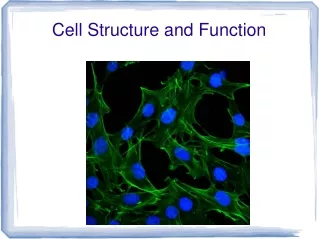
Microscopes are required to visualize cells • Types of microscopes • Light microscopes- compound light microscopes • Can resolve structures 200 nm apart • Electron microscopes • Can resolve structures 0.2 nm apart • 2 types • SEM (scanning electron microscope) • Forms 3-D images by beaming electrons onto the surface of the specimen • TEM (transmission electron microscope) • Also uses a beam of electrons that are transmitted through an ultra thin specimen, interacting with the specimen as it passes through it • The use of stains aids in viewing cell structure
All cells exhibit basic structural similarities • 1.) The genetic material is located in a centrally located nucleus or nucleoid. • 2.) A semi fluid matrix called cytoplasm fills the inside of the cell. • 3.) The plasma membrane encloses the cell and separates its contents from its surrounding.
Cell Organelles and Features Chapter 4 Structure of Lipid Bilayer
Cell Organelles and Features Chapter 4 Plasma Membrane- Fluid Mosaic Model • Membrane Proteins • Cell membranes often contain proteins embedded within the phospholipid bilayer.
Prokaryotic Cells • 2 types of Prokaryotes: Eubacteria and Archaea • Lack a membrane-bound nucleus • Instead genetic material is located in nucleoid • Prokaryotic cells possess • Cytoplasm • Plasma membrane • Cell wall • Ribosomes • However, they lack membrane-bound organelles. • Flagella are present in some prokaryotic cells • Used for locomotion • Rotary motion propels the cell
Prokaryotic cell walls • Function to protect the cell and maintain shape • Bacterial cell (Eubacteria) walls consist of peptidoglycan • May be gram + or gram – • Detected by gram-staining procedure • Gram +: thick, single layer peptidoglycan cell wall that retains the crystal violet stain (purple) • Gram -: more complex, multilayered cell wall that does not retain the crystal violet stain; stain with counterstain,safarin (pink) • Archaea lack peptiodoglycan
Eukaryotic cells • Possess a membrane-bound nucleus • More complex than prokaryotic cells • Compartmentalize cellular functions within organelles and endomembrane system • Organelles: membrane-bound structures that form compartments • Endomembrane system: system of connected membrane compartments • Have a cytoskeleton for support and to maintain cellular structure
Nucleus in eukaryotic cells • Stores the genetic material of the cell in multiple, linear chromosomes • DNA is organized with protein to form chromatin • Surrounded by a nuclear envelope • Composed of 2 phospholipid bilayers • Nuclear pores: opening in the nuclear envelope, allows proteins and nucleic acids in and out of the nucleus • Nucleolus (nucleoli): site of rRNA (ribosomal RNA) synthesis
Ribosomes • Site of protein synthesis in the cell • Composed of ribosomal RNA (rRNA) and proteins • Found within cytosol of the cytoplasm and attached to internal membranes • Each ribosome is composed of 2 subunits • Large and small subunit • Types of RNA • rRNA (ribosomal): provide a mechanism for decoding mRNA into amino acids and to interact with the tRNAs during translation • mRNA (messenger): carries coding information from DNA • tRNA (transfer): carries amino acids
Endomembrane system • Series of membranes throughout the cytoplasm • Divides the cell into compartments where different cellular functions occur • Endoplasmic reticulum (ER) • SER • RER • Golgi apparatus • Lysosomes
Rough endoplasmic reticulum • Membranes that create a network of channels throughout the cytoplasm • Attachment of ribosomes to the membrane gives a rough appearance • Synthesis of proteins to be secreted, sent to lysosomes or plasma membrane
Smooth endoplasmic reticulum • Relatively few ribosomes attached • Functions: • Synthesis of membrane lipids • Calcium storage • Detoxification of foreign materials
Golgi apparatus • Flattened stacks (golgi bodies) of interconnected membranes • Packaging and distribution of materials to different parts of the cell • Synthesis of cell wall components
Lysosomes • Membrane bound vesicles containing digestive enzymes to break down macromolecules • Destroy cells or foreign matter that the cell has engulfed by phagocytosis
Microbodies • Membrane bound vesicles • Contain enzymes • Not part of the endomembrane system • Types: • Glyoxysomes in plants contain enzymes for converting fats to carbohydrates • Peroxisomes contain oxidative enzymes and catalase
Vacuoles • Membrane-bound structures with various functions depending on the cell type • There are different types of vacuoles: • Central vacuole in plant cells • Contractile vacuole of some protists • Storage vacuoles
Mitochondria • Organelles present in all types of eukaryotic cells • Contain oxidative metabolism enzymes for transferring the energy within macromolecules to ATP • ‘Powerhouse’ of the cell • Surrounded by 2 membranes • Smooth outer membrane • Folded inner membrane with layers called cristae • matrix is within the inner membrane • Intermembrane space is located between the two membranes • Contain their own DNA
Plastids • Organelles like mitochondria found in plants that have a double membrane and contain own DNA • Examples: chloroplasts, chromoplasts, amyloplasts
Chloroplasts • Organelles present in cells of plants and some other eukaryotes • Contain chlorophyll for photosynthesis • Surrounded by 2 membranes • Inner and Outer membrane • Thylakoids are membranous sacs within the inner membrane • Grana are stacks of thylakoids
Endosymbiosis • Proposal that eukaryotic organelles evolved through a symbiotic relationship • One cell engulfed a 2nd cell and a symbiotic relationship developed • Mitochondria and chloroplasts are thought to have evolved this way • Evidence that supports • Mitochondria and chloroplasts: • Have 2 membranes • Possess DNA and ribosomes • Are about the size of a prokaryotic cell • Divide by a process similar to bacteria
Centrioles • Barrels of microtubules used in cell division
cytoskeleton • Network of protein fibers found in all eukaryotic cells • Support the shape of the cell • Keep organelles in fixed location • Helps in moving materials within the cell • Cytoskeleton fibers include • Actin filaments: responsible for cellular contractions, crawling, ‘pinching’ • Microtubules: provide organization to the cell and move materials within the cell • Intermediate filaments: provide structural stability
Cell movement • Cell movement takes different forms. • Crawling is accomplished via actin filaments and the protein myosin. • Flagella: undulate to propel the cell • Cilia: can be arranged in rows on the surface of a eukaryotic cell to propel the cell forward • Cilia and flagella have similar structure • 9-2 structure: 9 pairs of microtubules surrounded by a 2 central microtubules • Cilia are usually more numerous than flagella on a cell.
Extracellular structure • Includes • Cell walls in plants, fungi, some protists • Extracellular matrix surrounding animal cells • Cell walls • present surrounding the cells of plants, fungi, and some protists • the carbohydrates present in the cell wall vary depending on the cell type • plant and protist cell walls – cellulose • fungal cell walls - chitin
Extracellular matrix • ECM • surrounds animal cells • composed of glycoproteins and fibrous proteins such as collagen • may be connected to the cytoplasm via integrin proteins present in the plasma membrane