Download
1 / 27
270 likes | 333 Vues
Discover the intricate network of fibers in the cytoplasm, the function of microtubules, and the roles of vesicles and motor proteins. Learn about cilia, flagella, microfilaments, and the importance of cell walls in plants. Explore how tight junctions, desmosomes, and gap junctions play critical roles in cell interactions.

E N D
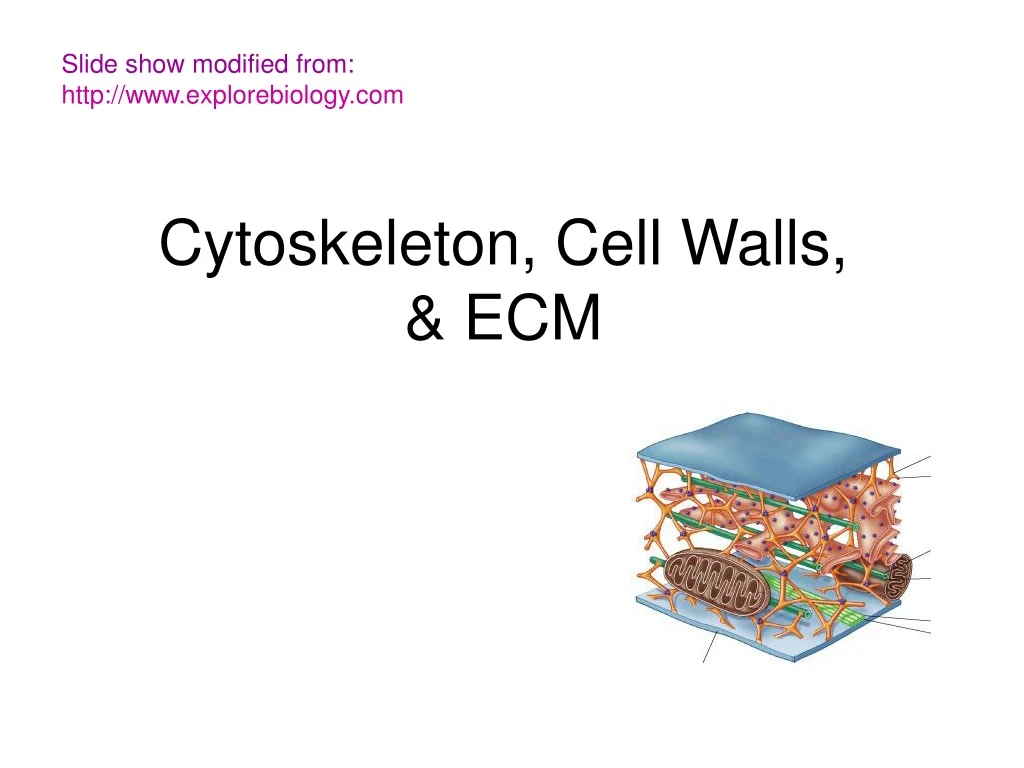
Slide show modified from:http://www.explorebiology.com Cytoskeleton, Cell Walls, & ECM
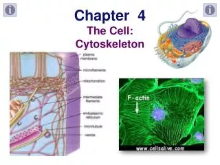
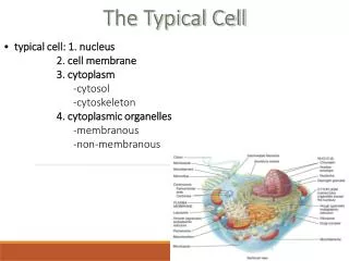
Cytoskeleton –network of fibers extending throughout the cytoplasm
MICROTUBULES FUNCTION Structural support and cell movement ~ Move chromosomes during cell division • Centrioles & spindle fibers ~ tracks guide motor proteins • Motor proteins: dynein & myosin ~ cell motility • Cilia & flagella
http://www.rpi.edu/dept/bcbp/molbiochem/MBWeb/mb2/part1/movies/kinesin.dcrhttp://www.rpi.edu/dept/bcbp/molbiochem/MBWeb/mb2/part1/movies/kinesin.dcr Vesicle Receptor for motor protein Motor protein (ATP powered) Microtubule of cytoskeleton MICROTUBULES Tracks guide motor proteins to destination (Motor proteins: dynein & myosin) ATP SEE MOTORPROTEINS inACTION • EXAMPLES • Vesicles containing neurotransmitters migrate to tips of nerve cells • Vesicles move to Golgi along cytoskeletal tracks • Cytoplasmic streaming http://python.rice.edu/~kolomeisky/transport.htm
Cilia and Flagella • Are locomotor appendages • Extensions of cytoskeleton Examples: Many unicellular protists move with flagella Some plant reproductive cells have flagella Cilia in oviducts move egg toward uterus Cilia lining windpipe sweep mucous out of lungs Flagellum in sperm cells (Prokaryotic flagella don’t have microtubules)
Outer microtubule doublet Plasma membrane 0.1 µm Dynein arms Central microtubule Outer doublets cross-linking proteins inside Microtubules Radial spoke Plasma membrane Basal body (b) 0.5 µm 0.1 µm Triplet Cross section of basal body Cilia and flagella share a common ultrastructure
FLAGELLUM (few, long) whip-like movement; cell moves in same direction as axis of flagellum CILIUM (many, short) oar-like movement; cell moves perpendicular to axis of cilium http://web.jjay.cuny.edu/~acarpi/NSC/13-cells.htm http://www.sk.lung.ca/content.cfm?edit_realword=hwbreathe
MICROFILAMENTS • STRUCTURE • Thinnest class of fibers • Twisted double chain of actin subunits ~ 7 nm in diameter • FUNCTION • Crosslinks with microtubules (cell shape) • Muscle cells: Actin filaments interact with myosin motor proteins to create muscle contraction • Amoeboid movement • Cytoplasmic streaming
Muscle cell Actin filament Myosin filament Myosin arm (a) Myosin motors in muscle cell contraction. MICROFILAMENTS Make up contractile apparatus of muscle • Contain the motor protein myosin in addition to actin
Cortex (outer cytoplasm): gel with actin network Inner cytoplasm: sol with actin subunits Extending pseudopodium Amoeboid movement • Actin filaments constantly form & dissolve making cytoplasm liquid or stiff during movement http://www.nextftp.com/jissen/ameba.gif
Nonmoving cytoplasm (gel) Chloroplast Streaming cytoplasm (sol) Parallel actin filaments Cell wall http://www.daviddarling.info/images/cytoplasmic_streaming.gif Cytoplasmic streaming • Speeds distribution of materials
Cell Walls of Plants Protection Maintain shape Also found in Prokaryotes, fungi, and some protists Composition varies with species/cell type Basic design: Microfibrils of polysaccharide cellulose embedded in matrix of other polysaccharides (like steel reinforced concrete)
Plant cell wall Structure PRIMARY CELL WALL MIDDLE LAMELLA- ~ between primary cell walls of adjacent cells ~ made of sticky polysaccharides (pectins) ~ glues cells together SECONDARY CELL WALL ~ built when cell stops growing ~ between plasma membrane and 1° cell wall
TIGHT JUNCTIONSMembranes of neighboring cells and pressed together & bound by proteins Forms continuous seal to prevent leakage of extracellular fluid across layer of cells DESMOSOMES (anchoring junctions) Act like “rivets” to fasten cells together into strong sheets Intermediate proteins (keratin) anchor desmosomes in cytoplasm GAP JUNCTIONS (communicating junctions) Channels connect to adjacent cells Special membrane proteins surround pore Necessary for communication between cells in heart muscle and animal embryos Types of intercellular junctions in animals
5 µm Figure 6.32 The Cell: A Living Unit Greater Than the Sum of Its Parts • Cells rely on the integration of structures and organelles in order to function