Download
1 / 22
220 likes | 248 Vues
Explore the intricate organization of the human skeleton, comprising 206 bones and divided into axial and appendicular portions. Learn about the skull's unique features, including the cranial and facial bones, and delve into the structure of the vertebral column with its series of vertebrae. Discover the functions and characteristics of each type of vertebrae, such as the cervical, thoracic, atlas, and axis. Gain insights into the critical role played by the hyoid bone and mandible, as well as the significance of the temporomandibular joint. Uncover the secrets of the human skeletal system in this comprehensive guide.

E N D
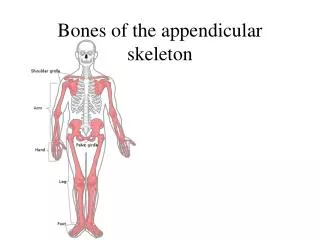
The Human Skeleton • 206 bones • Divided into two portions. • Axial Skeleton- Contains the bones which are in the mid-vertical axis of the body. • Appendicular Skeleton- Bones which are outside the mid-vertical axis, associated with the appendages.
Axial Skeleton • Skull • Contains 22 bones. • Bones of the skull include; (8) cranial, (13) facial and the (1) mandible. • Sutures- Connection between the bones of the skull. • Sinus-Chamber- Located in the skull, are lined with mucous membranes, and filled with air. • Connect with the nasal cavity to drain fluids and reduce the weight of the skull.
Axial Skeleton • Cranium (8) bones • 1.Frontal Bone- Large bone forming the anterior part of the skull (forehead). • Orbits: Eye sockets, partially formed by the frontal bone. • Supraorbital Foramen: Hole above each orbital where blood vessels pass.
Axial Skeleton • Cranium • 2.Parietial Bone(s)-Two bones which form most of the superior/lateral cranium. • Meet at the top of the skull at the sagittal suture. • Meet the frontal bone at the coronal suture.
Axial Skeleton • Cranium • 3.Occipital Bone- Thick bone which forms the posterior wall and floor of the cranium. • Meets the parietal bone at the lambdoid suture. • Foramen Magnum- Opening for the spinal cord. • Occipital Condyles- Processes that articulate with the first vertebrae, to allow head movement.
Axial Skeleton • Cranium • 4.Temporal Bone(s)- Two bones on either side of the cranium, below the parietal bones. • Meet the parietal bones at the squamous suture. • Styloid Process- Serves as an anchor for muscles of the tongue and pharynx.
Axial Skeleton • Cranium • 5. Sphenoid Bone- Butterfly shaped bone that forms the lower lateral walls and floor of the cranium, also the posterior walls of the orbits. • Optic foramen- Location of the optic nerve passage. • 6.Ethmoid Bone- Small bone anterior to the sphenoid bone. • Forms sections of the cranial floor, orbital walls, and nasal cavity.
Axial Skeleton • Facial Bones • Contains 13 immovable bones and a movable mandible. • 1.Maxillary Bone(s)- Two bones on each side of the face that form the upper jaw. • 2.Palatine Bone(s)- Two L-shaped bones that are posterior to the maxillary bones. • Forms posterior roof of mouth and floor of the nasal cavity.
Axial Skeleton • Facial Bones • 3.Zygomatic Bone(s)- Two bones on the side of the face that form the cheeks. • 4.Nasal Bone(s)- Two small rectangular bones that meet to form the bridge of the nose. • 5.Lacrimal Bone(s)- Two fingernail shaped bones, form part of the orbits medial walls.
Axial Skeleton • Facial Bones • 6.Vomer- Single bone along the midline within the nasal cavity. • Meets with the ethmoid bone to form the nasal septum, which divides the nasal cavity in half. • 7.Inferior Nasal Concha- Two thin scroll-like bones attached to the lateral walls of the nasal cavity. • Forms shelves which air is channeled into the nasal cavity.
Axial Skeleton • Facial bones • 8.Mandible-Single lower jaw bone, articulates with the temporal bones. • Only movable bone of the skull. • Forms TMJ (temporal-mandibular joint) • Lock Jaw • Dislocated Jaw
Axial Skeleton • Hyoid Bone • Single bone that doesn’t articulate with any other bones. • Located in the upper neck region. • Horse-shoe shaped • supports the tongue and provides attachments for muscles.
Axial Skeleton • Vertebral Column • Strong flexible rod that supports the trunk, while allowing for movement. • Extends from the base of the skull to the pelvis. • Comprised of a series of irregular bones known as vertebrae. • Between each vertebrae is an intervertebral disc. • The adult vertebral column contains 26 vertebrae (after fusion).
Axial Skeleton • Structure of typical vertebra: • Body: Thick, disc-shaped front portion, designed for supporting weight. • Vertebral Arch: Forms a ring to hold the spinal cord called the Vertebral Foramen. • Seven processes arise from this vertebral arch and serve for either joint formation or muscle attachment.
Axial Skeleton • Types of Vertebrae and their Structures. • Cervical- (7) Vertebrae of the neck region, the support the head. • The lightest of the vertebrae • Transverse foramen- small hole that permits the passage of arteries to the brain.
Axial Skeleton • Types of Vertebrae and their Structures. • Cervical- (7) Vertebrae of the neck region, the support the head. • Atlas- First vertebrae that connects with the occipital condyles of the cranium. • Contains no body • Allows for the up and down movement of the head • Axis- Second vertebrae that contains a tooth like projection called the: • Odontoid process- It projects up through the rings of the atlas and allows for twisting of the head.
Axial Skeleton • Types of Vertebrae and their Structures. • Thoracic- (12) Only vertebrae that articulate with the ribs. (upper and middle of the back) • Lumbar- (5) Vertebrae that are larger and thicker, this is due to the increase in the body weight stress they support. (lower back)
Axial Skeleton • Types of Vertebrae and their Structures. • Sacrum- Large triangular bone that forms the posterior part of the pelvis. • (5) vertebrae fused together. • Sacral Canal: contains the spinal cord. • Coccyx- A series of 3-5 fused bones that is attached to the sacrum by ligaments. (tail bone)
Axial Skeleton • Thoracic Cage • Formed by thoracic vertebrae, sternum, and the ribs. • Conical, basket-shaped structure (cone shaped). • Partially encloses the internal structures of the chest. • Supports the upper limbs.
Axial Skeleton • Thoracic Cage • Sternum- AKA breast bone, It’s a flat, narrow bone at the center of the chest. • Articulates with the clavicles on one end. • Articulates with the ribs via the costal cartilage. • Consists of three (3) parts: • Manubrium- Superior part • Body- Large middle part • Xiphoid Process- Small pointy inferior end.
Axial Skeleton • Thoracic Cage • Ribs • (12) sets in every individual. • Attach to the thoracic vertebrae in the back and the sternum in the front. • True Ribs- First seven (7) pair, connects directly to the sternum via the costal cartilage. • False Ribs- Remaining five (5) pair, have an indirect connection or no connection at all to the sternum. • Floating Ribs- Last two (2) or sometimes three (3) pairs of ribs, have no connection at all to the sternum.