Download
1 / 22
1.18k likes | 4.41k Vues
Single Photon Emission Computed Tomography (SPECT). Tom Walsh Dylan Schoo Sean Wilson Annie Moranski. History of Nuclear Medicine. Has it origins in a great many scientific discoveries dating back to the invention of the X-ray.

E N D
Single Photon Emission Computed Tomography (SPECT) • Tom Walsh • Dylan Schoo • Sean Wilson • Annie Moranski
History of Nuclear Medicine • Has it origins in a great many scientific discoveries dating back to the invention of the X-ray. • In 1946, radioactive iodine was used to treat a patient’s thyroid cancer. • By the 1950’s, radioactive nucleotides were being used to treat hyperthyroidism. • Soon after, it was possible to record snapshot images of form and structure or organs (liver, spleen, brain, gastrointestinal track, ect.).
History of Nuclear Medicine Cont. • By the 1980’s, nuclear medicine cameras and imaging computers allowed for the instillation of over 100 different procedures. • Nuclear medicine became an integral part of patient care, and an important diagnostic and therapeutic specialty.
History of SPECT • Edwards and Kuhl developed the MARK VI, the first Emission Computed Tomography (ECT) device. • MARK VI consisted of several sodium iodide photon detectors arranged in the rectangular shape around the head of the patient. • Tomomatic-32, first SPECT imaging device, was similar to the MARK VI but had 32 photon detectors.
History of SPECT Cont. • ECT was discredited due to unusable images. • Gained acceptance once X-ray CT image reconstruction algorithms were applied to ECT to take into account for attenuation for scatter in the body.
SPECT vs. PET • PET uses beta-plus-emitting radionuclides such as C-11, N-13, O-15, and F-18 which annihilate into two 511-keV photons that travel in opposite directions. • SPECT involves detection of gamma rays emitted singly from radionuclides such as Tc-99m, I-123, and In-111.
Pros of SPECT • Unlike MRI and X-ray, ECT produces 3-D images that relate an organ’s function. • This allows for better relay of extent of disease and reveals the course of the disease earlier. • Large amount of data on brain function already comes from SPECT scans. • Simple process with immediate results. • Much less expensive than MRI or PET ($1000 per scan) • Covered by insurance when brain injury is present.
Cons of SPECT • Unlike MRI and X-ray, there is an injection. • Claustrophobia is a cause for concern. • Quality of image can be lessened by patient movement.
Instrumentation • Radionuclides • Gamma emitting • 99mTc, 123I, 67Ga, 111In • Targeted for different tissues • attentuation • Collimator • Detector (scintillation) • Photomultiplier tubes • Circuitry • Preamplifier • Amplifier • Image Reconstruction
Gamma Cameras Scintillation type Gamma Cameras • Collimators • Lead (tungsten or platinum) • Excludes erroneous gamma rays (focus camera) • Spatial Resolution to Detection efficiency ratio
Gamma Cameras • NaI(Tl) crystals • Produce lower frequency photons • Photomultiplier tubes • Transform photons to electrical signals Using photocathode
Gamma Cameras Multiple Camera Systems • Reduce acquisition time • Rotating camera vs. full ring camera
Image Reconstruction • Gamma rays counted into matrix • Filtering • Convolution Method (9 point smoothing) • Fourier Method • Digital vs. Annalog • Too few pixels • Too few bytes per pixel
Technological Advances • Problems with SPECT: • Long scan times • Low resolution • Attenuation • Solutions: • Triple headed cameras drastically reduce scan times • Improved cameras and computers enhance resolution • Visual Tracking Systems to monitor patient movement and correct images accordingly • Attenuation Correction software
Clinical Applications • In the 70’s & 80’s, SPECT was largely replaced by CAT and MRI scans because they provided superior resolution. • Recently, SPECT has returned to prominent use, especially in diagnosing cardiac and neurological abnormalities. • While CAT and MRI scans only provide images of static brain anatomy, SPECT offers clues to brain function by tracing blood allocation.
Diagnosis of Neurological and Psychiatric Disorders with SPECT • Doctors study two kinds of brain SPECT images: Surface Image: Active Image: Full symmetrical activity across cortical surface High activity in cerebellum and visual or occipital cortex • Doctors look for too much/little activity in a certain area, or asymmetry in areas that should be symmetrical.
SPECT Images of Common Neurological and Psychiatric Disorders Alzheimer’s Disease pervasive hypoperfusion Right Sided Stroke Depression increased limbic activity (left) and decreased prefrontal and temporal lobe activity Head Trauma to left PFC - severe aggression problems/violence
SPECT Images of Common Neurological and Psychiatric Disorders ADHD Normally (left) and while performing a concentration task (right) Schizophrenia Before (left) and after (right) medication Alcohol Marijuana Cocaine Heroin
Cardiac SPECT Images Normal Myocardial Perfusion at stress and rest Single Vessel Coronary Artery Disease at stress and rest SPECT image shows limited perfusion (horseshoe shape rather than donut) especially under stress. SPECT image exhibits full perfusion (bright, donut shape) both at rest and under stress
Current studies using SPECT • Alcohol induced brain activity • ADHD • SPECT and aggressiveness • ADD
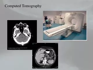
CT and SPECT SPECT provides functional information, but it has relatively poor spatial resolution and can lack the anatomical information needed to localize or stage disease. CT offers excellent spatial resolution and rich anatomical detail.