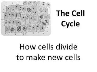
The cell cycle
The cell cycle The cell cycle The life of a cell from its formation to its division The cell cycle The big picture A walk through the cell cycle Control of the cell cycle Cancer: a lack of control of the cell cycle The big picture “Every cell from a cell”-Rudolf Virchow

The cell cycle
E N D
Presentation Transcript
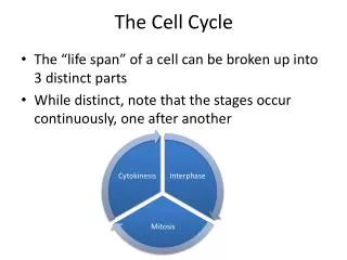
The cell cycle The life of a cell from its formation to its division
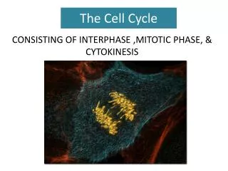
The cell cycle • The big picture • A walk through the cell cycle • Control of the cell cycle • Cancer: a lack of control of the cell cycle
The big picture • “Every cell from a cell”-Rudolf Virchow • The continuity of life is based on reproduction of cells, cell division • Cell division results in genetically identical daughter cells • Unicellular organisms use cell division as a means of reproduction (asexual reproduction) • Sexually reproducing organisms develop from a fertilized egg via cell division • In multicellular organisms, cell division is critical to growth and repair
A walk through the cell cycle What happens? • DNA, which is arranged in chromosomes, duplicates • Each daughter cell receives the full genome • Cytoplasm divides
Cell wall Origin of replication Plasma membrane E. coli cell Bacterial chromosome Two copies of origin Origin Origin A walk through the cell cycle: Prokaryotes • Cell division begins when the chromosome begins to divide • One copy moves to one pole, the other copy moves to other end of the cell • The cell elongates • Replication of the DNA is completed, the plasma membrane moves inward and the cell wall develops • This is asexual reproduction (binary fission)
Cell wall Origin of replication Plasma membrane E. coli cell Bacterial chromosome Two copies of origin Origin Origin A walk through the cell cycle: Prokaryotes This process is more complicated in eukaryotes • Cell division begins when the chromosome begins to divide • One copy moves to one pole, the other copy moves to other end of the cell • The cell elongates • Replication of the DNA is completed, the plasma membrane moves inward and the cell wall develops • This is asexual reproduction (binary fission)
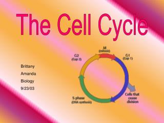
INTERPHASE S (DNA synthesis) G1 Cytokinesis G2 Mitosis MITOTIC (M) PHASE A walk through the cell cycle: Eukaryotes • The cell cycle involves: • Interphase • Growth and DNA duplication • M-phase • Mitosis-division of the nucleus • Cytokinesis-division of the cytoplasm DNA molecules
INTERPHASE S (DNA synthesis) G1 Cytokinesis G2 Mitosis MITOTIC (M) PHASE A walk through the cell cycle: Eukaryotes • The cell cycle involves: • Interphase • Growth and DNA duplication • M-phase • Mitosis-division of the nucleus • Cytokinesis-division of the cytoplasm DNA molecules Let’s watch a movie about this!
A walk through the cell cycle: Duplication of genetic information during eukaryotic cell division Interphase: • The cell grows during G1 • The chromosomes are duplicated during S phase • Unlike proks, they have many! • When the cell is not dividing (even as it duplicates its DNA) the chromosomes are long and thin • After duplication the chromosomes are shorter and thicker • Each duplicated chromosome has two identical sister chromatids (attached at the centromere) • Each will become a chromosome in the new daughter cells 0.5 µm Chromosomes DNA molecules Chromosome duplication (including DNA synthesis) Centromere Separation of sister chromatids Sister chromatids Centromere
Metaphase Anaphase Telophase and Cytokinesis G2 of Interphase Prophase Prometaphase Centrosomes (with centriole pairs) Early mitotic spindle Centromere Chromatin (duplicated) Fragments of nuclear envelope Nonkinetochore microtubules Aster Cleavage furrow Metaphase plate Nucleolus forming Daughter chromosomes Nuclear envelope forming Centrosome at one spindle pole Spindle Nuclear envelope Kinetochore Chromosome, consisting of two sister chromatids Kinetochore microtubule Plasma membrane Nucleolus A walk through the cell cycle: G2 phase (Interphase) During the G2 phase of interphase: -Nuclear envelope is still intact (nucleoli are still visible) -Centrosomes have formed -Duplicated chromosomes cannot be seen
Metaphase Anaphase Telophase and Cytokinesis G2 of Interphase Prophase Prometaphase Centrosomes (with centriole pairs) Early mitotic spindle Centromere Chromatin (duplicated) Fragments of nuclear envelope Nonkinetochore microtubules Aster Cleavage furrow Metaphase plate Nucleolus forming Daughter chromosomes Nuclear envelope forming Centrosome at one spindle pole Spindle Nuclear envelope Kinetochore Chromosome, consisting of two sister chromatids Kinetochore microtubule Plasma membrane Nucleolus A walk through the cell cycle: Mitosis Prophase: -Chromosomes condense and are visible (nucleoli disappear) -Mitotic spindle begins to form (contains centrosomes, aster, and microtubules) -Centrosomes move away from each other
Metaphase Anaphase Telophase and Cytokinesis G2 of Interphase Prophase Prometaphase Centrosomes (with centriole pairs) Early mitotic spindle Centromere Chromatin (duplicated) Fragments of nuclear envelope Nonkinetochore microtubules Aster Cleavage furrow Metaphase plate Nucleolus forming Daughter chromosomes Nuclear envelope forming Centrosome at one spindle pole Spindle Nuclear envelope Kinetochore Chromosome, consisting of two sister chromatids Kinetochore microtubule Plasma membrane Nucleolus A walk through the cell cycle: Mitosis Prometaphase: -The nuclear envelope fragments -Microtubules attach to the chromatids (at the kinetochores) -Chromosomes are jerked back and forth -Other microtubules interact (connecting from different poles)
Metaphase Anaphase Telophase and Cytokinesis G2 of Interphase Prophase Prometaphase Centrosomes (with centriole pairs) Early mitotic spindle Centromere Chromatin (duplicated) Fragments of nuclear envelope Nonkinetochore microtubules Aster Cleavage furrow Metaphase plate Nucleolus forming Daughter chromosomes Nuclear envelope forming Centrosome at one spindle pole Spindle Nuclear envelope Kinetochore Chromosome, consisting of two sister chromatids Kinetochore microtubule Plasma membrane Nucleolus A walk through the cell cycle: Mitosis Metaphase: -Centrosomes are now at opposite poles -Chromosomes are lined up on the metaphase plate -All chromosomes are attached to each of the poles
Metaphase Anaphase Telophase and Cytokinesis G2 of Interphase Prophase Prometaphase Centrosomes (with centriole pairs) Early mitotic spindle Centromere Chromatin (duplicated) Fragments of nuclear envelope Nonkinetochore microtubules Aster Cleavage furrow Metaphase plate Nucleolus forming Daughter chromosomes Nuclear envelope forming Centrosome at one spindle pole Spindle Nuclear envelope Kinetochore Chromosome, consisting of two sister chromatids Kinetochore microtubule Plasma membrane Nucleolus A walk through the cell cycle: Mitosis Anaphase: -The connection between chromatids at the centromere is cleaved (each chromatid is now a chromosome) -Chromosomes are pulled to opposite poles -The cell elongates -Anaphase ends when the chromosomes reach the poles
Metaphase Anaphase Telophase and Cytokinesis G2 of Interphase Prophase Prometaphase Centrosomes (with centriole pairs) Early mitotic spindle Centromere Chromatin (duplicated) Fragments of nuclear envelope Nonkinetochore microtubules Aster Cleavage furrow Metaphase plate Nucleolus forming Daughter chromosomes Nuclear envelope forming Centrosome at one spindle pole Spindle Nuclear envelope Kinetochore Chromosome, consisting of two sister chromatids Kinetochore microtubule Plasma membrane Nucleolus A walk through the cell cycle: Mitosis Telophase: -Two daughter nuclei form in the cell (nuclear envelope forms and nucleoli appear) -Chromosomes become less dense -Mitosis is complete
100 µm Cleavage furrow Daughter cells Contractile ring of microfilaments (a) Cleavage of an animal cell (SEM) A walk through the cell cycle: Cytokinesis Cytokinesis -Usually occurs during late telophase -In animal cells, it occurs by cleavage -A cleavage furrow forms -Actin microfilaments and myosin motor proteins cause it to contract
Vesicles forming cell plate Wall of parent cell 1 µm Cell plate New cell wall Daughter cells (b) Cell plate formation in a plant cell (TEM) A walk through the cell cycle: Cytokinesis Cytokinesis -Usually occurs during late telophase -In plant cells, the cell wall must be constructed -cell plate is formed -it develops into a cell wall
INTERPHASE S (DNA synthesis) G1 Cytokinesis G2 Mitosis MITOTIC (M) PHASE A walk through the cell cycle: In review • The cell cycle involves: • Interphase • Growth and DNA duplication • M-phase • Mitosis-division of the nucleus • Cytokinesis-division of the cytoplasm DNA molecules
Control of the cell cycle • Some cells divide more than others • Timing and rate of cell division are highly regulated
Control of the cell cycle • Some cells divide more than others • Timing and rate of cell division are highly regulated How is the cell cycle regulated?
Control of the cell cycle Evidence for cytoplasmic signals (Johnson and Rao 1970) EXPERIMENT Experiment 1 Experiment 2 G1 S G1 M RESULTS M S S M
5 30 4 20 3 % of dividing cells (– ) Protein kinase activity (– ) 2 10 1 0 0 400 100 200 300 500 Time (min) Control of the cell cycle Protein kinases- enzymes that can activate or inactivate other proteins by phosphorylating them (Moreno et al. 1989)
Control of the cell cycle G1 checkpoint The sequential events of the cell cycle are directed by a distinct cell cycle control system • There are checkpoints where the cell cycle stops until a go-ahead signal is received (cyclin-dependent kinases are involved with these check points) • Ie. If the cell does not get the go ahead at the G1 check point, it will enter a non-dividing phase (G0) Control system S G1 G2 M M checkpoint G2 checkpoint
Control of the cell cycle G1 checkpoint The sequential events of the cell cycle are directed by a distinct cell cycle control system • There are checkpoints where the cell cycle stops until a go-ahead signal is received (cyclin-dependent kinases are involved with these check points) • Ie. If the cell does not get the go ahead at the G1 check point, it will enter a non-dividing phase (G0) • Ie. M check point involves the connection of all chromosomes to the spindle (internal control) Control system S G1 G2 M M checkpoint G2 checkpoint
Control of the cell cycle Anchorage dependence External signals • Cell may not divide if an essential nutrient is lacking • Growth factors (molecules released from other cells or tissues can stimulate division) • Density-dependent inhibition-crowded cells stop dividing • Anchorage dependence-to divide cells must be attached to a substrate Density-dependent inhibition Density-dependent inhibition 25 µm 25 µm (b) Cancer cells (a) Normal mammalian cells
Control of the cell cycle Anchorage dependence External signals • Cell may not divide if an essential nutrient is lacking • Growth factors (molecules released from other cells or tissues can stimulate division) • Density-dependent inhibition-crowded cells stop dividing • Anchorage dependence-to divide cells must be attached to a substrate Density-dependent inhibition Density-dependent inhibition 25 µm 25 µm (b) Cancer cells (a) Normal mammalian cells Cancer cells do not exhibit density-dependent inhibition or anchoring dependence
Cancer: A loss of control of the cell cycle • Cancer cells divide excessively invading other tissues, they can lead to death • Why? • Some make their own growth factors • Some can divide without growth factors • They lack density-dependent inhibition and anchorage dependence
Cancer: A loss of control of the cell cycle • The disease begins with transformation-the process of converting a normal cell to a cancer cell • If the transformed cell avoids the immune system it may proliferate and form a tumor • A tumor can be benign or malignant (invasive enough to impair other organs) • Cells in malignant tumors may have: • Unusual chromosome numbers • Changes on the cell surface that allow them to spread • Cause blood vessels to grow near them
You should understand: • The importance of cell division • The phases of the cell cycle: interphase, mitosis, and cytokinesis • Factors that control the cell cycle: molecules involved, checkpoints, internal and external controls • Cancer cells have lost control of the cell cycle The cell cycle