Download
1 / 48
480 likes | 859 Vues
Protocols of conservative mangement of condyle neck fracture in children . 가천의과대학 부속 길병원 치과센터 구강악안면외과 김현민. 차례. 서론 본론 – 소아과두골절의 특징 소아과두골절의 치료 case report 1 2 결론 . 서론 ( 개념의 변화 ). 소아 하악 과두골절 – 악기능 , 안면성장장애를 줄 수 있다 치료방법 – 2-30 년전 ; 관혈적 정복술

E N D
Protocols of conservative mangement of condyle neck fracture in children 가천의과대학 부속 길병원 치과센터 구강악안면외과 김현민
차례 • 서론 • 본론 –소아과두골절의 특징 소아과두골절의 치료 case report 1 2 • 결론
서론(개념의 변화) • 소아 하악 과두골절 –악기능, 안면성장장애를 줄 수 있다 • 치료방법 – 2-30년전;관혈적 정복술 최근;보존적 치료(장기 추적연구 결과, 악골성장과 기능회복에 뛰어난 성과) • *Dahlstrom-15년간 추적조사 (대부분의 환자에서 주요 성장장애나 기능이상은 없었으나 10세 이후 골절시 성장장애나 기능이상 발생율이 높다) • *Norholt etc – (보존적 치료가 10세 이하나 10대 초기의 어린이에서 장애나 이상을 덜 발생)
서론(개념의 미확립) • *Giuus-Moe (총 43명을 보존적 치료시 14명 편위 발생) • *Lund (27명중 6명 하악골 성장장애 또는 과성장) 현재- 보존적 치료가 만족스러운 결과를 가져오지만 성장장애, 기능장애, 안면비대칭, 악관절증을 일으키는 예등 예측할 수 없는 요소가 있다.
본론(예측을 위한 요소) 1. 소아과두골절 형태 2. 특정 연령 3. 치료방법(보존적치료) - soft diet/악간고정기간 - instruction,exercises,observation - orthodontic aftercare - surgery • 보존적 치료 후 발생되는 성장장애, 기능장애 등 예측 못하는 후유증을 최소로 하기 위해서 상기 요소에 대한 이해가 중요하리라 보며 이들에 대한 문헌을 돌이켜 보고자 한다.
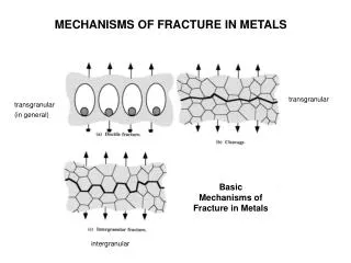
소아과두골절의 특징 1. 어른에서 악관절내장증이 주로 발생하는 반면 악관절 유착이 잘 생김.(특히 관절낭내 골절시) -관절강내로 혈액공급이 잘 되 골형성 물질을 채우기 때문 2.골절의 위치와 종류, 손상 받은 당시의 나이 , 악간 고정기간, 관절원판의 손상,손상 후 과두의 골절편과 관절와의 관계등에 영향 3.골절보다 붕괴되기 쉽다. Crush injury가 많다. -소아 과두가 피질골이 얇고 과두경부가 넓다 -subarticular layer의 혈액공급이 풍부
하악과두의 성인과 소아의 차이점구강악안면외과학 교과서 인용
소아과두골절의 특징 4.10세전에 유착이 잘. 5.관절자체내 골유합 및 섬유성 유합에 의한 true ankylosis, 연조직 손상에 의한 섬유화나 반흔형성등에 의한 pseudoankylosis 유발 6.10세 이하, 관절낭내 골절과 골절편이 변위된 경우, 복합 분쇄골절된 경우, 근돌기와 관골 골절이 동반될 경우 유착 가능성 증가되므로 짧은 고정기간, 술후 물리치료와 장기 관찰등이 예방책
소아과두골절의 complications • 하악골 개구시 변위 • 안면비대칭 • 일시적 안면신경 마비 • 하악골 운동시 악관절 동통 • 악관절 잡음, 감염, 과두 흡수
소아과두골절의 치료(문제의 제시) 1)surgical ; nonsurgical 2)post op methods IMF ; non IMF appliance ; physical exercise 3)F/U 성장장애, 기능장애, 합병증에 대한 치료 fixed ortho tx ; surgical intervention
문제를 풀어봅시다!!! 1번 문제 Surgical (open) ; nonsurgical(cloed) #치료의 제시 ; 소아 환자의 condyle neck fx시 deviation이 심하더라도 최대한 conservative 하여야 한다. 단, 골절이 심할 수록 IMF를 시행하여 occlusion을 확립하고 appliance를 적용하여야 한다. 다른 부위와 동반된 경우는 surgical tx를 고려하여야 한다.
Fractures of Mn CondyleBy James R. Haywasrd etc J. Oral Maxillofac Surg 51:57-61.1993 • Closed : open reduction에 미치는 요인들 1)age of pt 2)level of fx 3)degee of displacement 4)direction of displacement 5)medical status of pt 6)concomitant injures 7)status of presence dentition 8)ease in establishing adequate occlusion 9)presence of foreign body
Possible decision tree for condylar fractures 1)absolute indications for oepn reduction - 중두개와로 전위 - CR 적절한 교합을 얻가 어려울때 - lat. Capsular displacement - 관절장내 이물(eg.Gunshot wound)
2)relative indications - 중안면부 분쇄골절에 대한 견고고정이 불가능한 경우 양측성과두골절 - 전신적 질환으로 splinting이 추천되지 않는 양측 또는 단독 과두골절 (eg, seizure disorders,alcoholism) - rigde atrophy로 인한 splint 되기 힘든 양측성 과두골절 - gnathologic 문제를 야기하는 양측성 과두골절 (eg, unstable occlusion)
Long-term evaluations of pediatric condyle fracture patients (By 오승환. 대한악안면성형재건외과학회 vol 24,# 6, 2002) 1)소아의 하악과두골절의 보존적 치료후 악관절 기능장애 및 성장장애의 발생률은 약20%-25%로 매우 높았다. 2)7-10세 사이의 연령에서 악관절 기능장애와 성장장애가 가장 컸다.(2차 성장기를 앞두고 있는 나이) 3)손상부위가 하악과두에 가까울수록 악관절기능장애와 성장장애가 컸다.
문제를 풀어봅시다 *2번 문제 소아과두골절의 치료방법? 1)soft diet + immediate mobilization (open bite-elastics guide) 2)straight movement + IMF(2주) + guide elastics + orthodontic tx ( 초기 functional + 마지막 fixed appliance )
3) 부가적인 IMF 없이 4-6 개월간 activator (myofunctional appliance) 16 시간이상 장착 4) 5-10일간 elastic traction으로 immobilization + elastic guide 하 PT + 최소 1년간 closed observation (functional appliance)
치료방법1 Long term results of nonsugical management of condylar fx in children (By J. Hovinga etc Int J. Oral Maxillofac Surg 1999; 28;429-440) 1)대상 : 28 area 25 pt 2)치료방법 ; 1)enhance straight opening & closing movement 2)2주간 IMF- guide elastics 3)orthodontic tx- 초기 functional appliance + fixed appliance 3)결론 ; High condyle fx- good regeneration 양상 Low condylar & intracapsular fx- facial asymmetry 양상 증가 (malocclusion-orthosurgery need)
치료방법2 Condylar process fx in children; a follow up study of fx with total dislocation of the condyle from the glenoid fossa (By Hanna Thoren JOMS 59 ; 768-773,2001) 1)대상 ; 15세 이하 34명 2)치료방법 ; soft diet, early mobilization 초기 개교합시 mobilization 을 허용하면서 guide elastics 사용 3)결론 ; TMJ56%symptoms,72%sign X-ray 76.5%incomplete remodelling, 64.7%mn asymmetry 소아에서 발생된 변위성 과두돌기 치료시 conservative tx는 방사선적인 면에서 나쁜 결과를 가져왔지만 악골의 기능적인 면에서 장기간 만족할 만한 결과를 가져왔다.
치료방법3 Conservative tx of unilateral condylar fx in children; a long term clinical & radiologic F/U of 55 pt (By H. Strobl etc Int J Oral Maxillofac Surg 1999; 28;95-98) 1)대상; 2.5-9.8세 55명 2)치료방법; nonsurgical-functional way using an intraoral myofunctional appliance 이용 6,12,24,48,72주 F/U 3)방법; 부가적인 IMF 없이 4-6개월간 activator를 최소 16시간이상 장착
3)결론 ;48주에 교합, 기능장애, TMJ장애 소실되었고 remodeling 완전해짐 4)2-6세 47명 – no,only slight deformity 7-10세 8명 – 2 moderate ,4 hypertrophic deformity, 2 neck height 감소 5)Displaced & dislocated condyle fx 시 condylar remodeling이 나타난다.
장치의 적용 1 Functional therapy in hemifacial microsomia / therapeutic protocol for growing children (By alessandro silvestri etc, J Oral Maxillofac Surg 54;271-278,1996) HM를 보이는 성장아동에서 AFA(asymmetrical functional activator)를 이용은 조화로운 상하악성장을 유도한다. 그러나 심각한 형태일 경우 고정성 교정치료와 surgical intervention이 요구된다.
성공예/ skeletal :1) small Mn with normal shape 2)condyle, ramus,sigmoid notch identifiable but grossly distorted; Mn strikngly different in size & shape from normal 3)Minimal & moderate soft tissue contour defect with no cranial nerve involvement
장치의 적용2 Use of activator appliances in pediatric pt treated with costochondral grafts for TMJ ankylosis; anaylsis of 13 cases (By Hossein Behnia etc J Oral Maxillofac Surg 55;1408-1414,1997) 1) 대상; 장기간 과두 ankylosis로 인한 안면비대칭과 occlusal canting을 보인 소아 2) 방법; condylectomy & immediate costochondral rib graft reconstruction +activator를 대칭성과 교합이 유지 될 때까지 장기간 적용
3) 결론 ; activator를 이용한 functional therapy가 적용되지 않을 경우 하악 변위, 교합 부조화 및 비대칭,그리고 TMJ reankylosis와 같은 합병증이 발생하였다.
Case report 1 • Pt ; 장동현(4/M) • Hx; TA • Dx; Mn fx on Rt parasym & Lt subcondyle • Tx;1)2002.12.13 interdental wiring 2)2002.12.18 circumferential wiring under G/A on Rt parasym & closed reduction on Lt subcondyle
3)soft diet & occlusion observation - inf, 교합이상 여부 closed observation - physical exercise-2주 4)post op 약 4주 wire removal
Case report 2 • Pt ; 최은진 (4/F) • Hx ; 2002.6.10 계단 slip down • Pi ; facial open lac + bony mobildity & deviation • Dx ; Mn fx on Rt body & Lt subcondyle • Tx ; 1)2002.6.10 circumferential wiring reduction & fixation
2)soft diet & mouth opening limit 3)post op 3주 wire removal
Conclusions • 소아과두골절은 심미적 기능적 문제를 일으킬 가능성이 높다 –초기 교합의 형성+ 기능적 치료(Frankel,Activtor)의 적용 + 주기적 검사(특히 post truma 4주) • 나이 ; 2-6세 아동에서는 보전적 치료로 인한 효과가 좋다 7-10세 아동에서는 보전적 치료의 효과가 이전 나이보다 떨어지며 합병증 가능성이 높아진다.
Conclusions • 치료방법의 변화 –유아 (immediate mobilization methods); 1)soft diet & early mobilization 2)close observation of occlusion 3)open bite시 elastics guiding
Conclusions 치료방법의 제시 ; 소아 1)5-10일immobilization & elastic guide 2)elastic guide하에 전, 측방, 개구 신장운동 –보호자 physical therapy 3)과두재형성과 근골격근 조화를 위한 functional appliance로 하악 projection 과 대칭성 확립
Conclusions • 치료방법의 제시- 골절 방식 1)불완전 형태 골절, 높은 위치 골절-주기적 관찰 2)골절편의 변위가 심하거나 낮은 위치에서 발생한 과두골절 변위가 심한 경우-관혈적 정복술 / IMF(arch bar, wire,splint) + elastic move
마지막 • 소아의 하악 과두 골절 환자를 치료시 7-10세 연령대에서 발생한 경우, 손상이 하악 과두에 가까이 손상이 있을 경우, 골절편의 변위가 심한 경우 기능장애나 성장장애가 심하므로 보다 적극적으로 보존적 치료에 임해야. • 소아과두골절 치료는 시기 적절한 하악골의 기능적 운동에 의한 하악골 과두의 재형성을 유도하여 수술 후 장기간에 걸쳐 주기적인 관찰을 요한다.