CELL PHYSIOLOGY-----2
LECTURE----3 DR ZAHOOR ALI SHAIKH. CELL PHYSIOLOGY-----2. CYTOPLASM CYTOSOL AND CYTOSKELETON. What is Cytosol ? Cytosol is semi liquid portion of the cytoplasm that surrounds the organelles. It occupies 55% of cell volume. It is like a gelatinous mass. FUNCTIONS OF CYTOSOL.

CELL PHYSIOLOGY-----2
E N D
Presentation Transcript
LECTURE----3 DR ZAHOOR ALI SHAIKH CELL PHYSIOLOGY-----2
CYTOPLASM CYTOSOL AND CYTOSKELETON • What is Cytosol ? • Cytosol is semi liquid portion of the cytoplasm that surrounds the organelles. It occupies 55% of cell volume. • It is like a gelatinous mass.
FUNCTIONS OF CYTOSOL 1- Ribosomal protein synthesis Free ribosome's are present in cytosol , which synthesize protein. 2- Intermediary Metabolism It involves degradation, synthesis. Thousands of Enzymes involved in Glycolysis are found in cytosol.
FUNCTION OF CYTOSOL (CONT) 3- Storage of Fat, carbohydrate (Glycogen), and secretary vesicles • These stored material(fat and glycogen) are known as Inclusions. • Glycogen storage-Maximum in liver and muscle cells. • Fat storage-Maximum in Adipose tissue cells.
CYTOSOL (CONT) • When food not supplied , stored glycogen and fat in cytosol are broken down to release glucose and fatty acids. • Within Cytosol is Cytoskeleton-that gives shape to cell and responsible for various cell movements.
CYTOSKELETON IN CYTOPLASM • Cytoskeleton is complex Protein Network present in the cytoplasm. This network has 1- Microtubules 2- Microfilaments 3- Intermediate Filaments
CYTOSKELETON (CONT) 1) MICROTUBULES • It is protein in nature, maintains the shape of the cell. • Microtubules play important role in movement of cilia and flagella.
CYTOSKELETON (CONT) Note • Cilia - tiny hair like protrusions. Ciliated cell are found in Respiratory Tract, oviduct of female reproductive tract. • Flagella - only Human cell that have flagella are sperm. Motion of flagellum or tail enables sperm to move.
CYTOSKELETON (CONT) 2). MICROFILAMENTS • They are important to the cell for a) Contractile systems E.g. Actin and Myosin are (protein molecule) present in muscle for muscle contraction • b) Mechanical stiffness of cell E.g. in Microvilli (Non-motile ,hair like projection from the surface of epithelial cells of small intestine)
CYTOSKELETON (CONT) • Both Microtubules and Microfilaments form stable structures. • Example: 1. Cilia, Flagella by Microtubules. 2. Actin and myosin (muscle contractile units) and microvilli by Microfilaments.
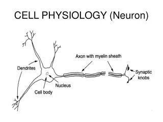
CYTOSKELETON (CONT) 3). Intermediate Filament • They are intermediate in size between microtubules and microfilaments [7 – 11 nm in diameter] – hence their name. • Examples – Neurofilaments are intermediate filament present in nerve cell. • Present in skin as keratin protein.
PLASMA MEMBRANE • Plasma membrane covers the cell. It is made of LIPIDS and PROTEIN. • It is thin can not be seen by light microscope but with electron microscope. • It separates the cell from extracellular fluid. • Exchange of material takes place across the plasma membrane.
Plasma Membrane (cont) • It is selectively permeable. • Maintains difference in Ion concentration between interior and exterior of the cell. • These ionic difference(ions have charge) are important for electrical activity of the cell.
Plasma Membrane(cont) • Cells have Membrane Potential—it is negative inside. • Nerve and Muscle cell has the ability to change their potential on stimulation.
Plasma Membrane Structure • Plasma membrane is a fluid lipid bi-layer embedded with protein . • Plasma membrane has mostly lipids and protein plus small amount of carbohydrate. • Membrane carbohydrate is at the surface forming glycoprotein and glycolipids.
Plasma Membrane Structure (cont)1. LIPID LAYER • 1. Lipid layer (mostly phospholipids and little cholesterol) is not rigid but flexible and fluid in nature. • Fluid nature of cell membrane permits it to change the shape e.g. Red blood cells can change the shape while passing through capillaries.
Plasma Membrane (cont) • Lipid layer– Three important functions 1. Phospholipids forms basic structure of membrane. 2. It is Hydrophilic (water liking) outside and Hydrophobic (water fearing) inside. 3. It is responsible for fluid nature of cell membrane.
Plasma Membrane (cont)2. Membrane Protein • 2. Membrane Protein Functions • Different protein in the plasma membrane have different specialized functions. i. Protein form channels– Ions pass through these channels, e.g. Na+, K+, Ca++. ii. Channel allow up to the size of 0.8nm (Nanometer).
Plasma Membrane Protein Function (cont) iii. Other protein serve as carrier molecule. Each carrier can carry only particular molecule e.g. Thyroid gland cells can carry Iodine from blood to Thyroid gland (other body cells can not). iv. Protein on cell membrane (outer surface) serve as receptor sites e.g. Hormones can act on different receptors.
Plasma Membrane protein (cont) v. Some protein on the interior of the cell combine with secretary vesicles and empty its content outside by EXOCYTOSIS.
Plasma Membrane protein (cont) vi. Other protein function as enzymes on both sides of membrane (they are embedded in the plasma membrane). vii. Protein on plasma membrane serve as CELL ADHESION MOLECULES (CAMs). viii. 0ther protein with carbohydrate are important to recognize “SELF”.
3. Plasma Membrane Carbohydrate - Functions i. Work as self identity marker. ii.Carbohydrate surface marker are involved in Tissue growth within physiological limits.
Cell to Cell Adhesions • Cells are held together by THREE different ways: 1. Cell Adhesions Molecules (CAM) —proteinin plasma membrane 2. Extracellular Matrix 3. Specialized Cell Junctions
Cell to Cell Adhesions (cont) 1. Cell Adhesions Molecules (CAMs) already mentioned (made by proteins).
Cell to Cell Adhesions (cont) 2. Extra cellular Matrix (ECM) - It works as biological “Glue” - It is mesh work of fibrous protein embedded in watery gel substance of complex carbohydrate.
Cell to Cell Adhesions (cont) • ECM Present in this gel (carbohydrate) are THREE types of protein fibers: i. Collagen e.g. skin , gums ii. Elastin- rubber like protein, present in LUNGS –stretch and recoil during breathing iii. Fibronectin- increases cell adhesion , it is decreased in cancer cells , therefore cancer cell break and metastasize (spread in body).
Cell Adhesion (cont) • Useful information • ECM ( extra cellular matrix ) comes from where ? • It is secreted by Fibroblast present in the matrix. • In some tissues ECM becomes highly specialized to form structures like cartilage and tendons or upon calcification hardened structure of Bone and Teeth.
3) Specialized Cell Junctions • In addition to cell adhesion molecules (CAM) and extra cellular matrix (ECM) , some cells are connected by CELL JUNCTIONS. • CELL JUNCTIONS THREE TYPES: 1) Desmosomes 2) Tight junctions 3) Gap junctions
CELL JUNCTIONS (cont) • Desmosomes • Desmosome consist of cytoplasmic thickening on the inner side of the of each of the two adjacent cells—called PLAQUE. • Strong glycoprotein filaments are attached to plaque on both sides. • Desmosome are present through out the body but more in tissues where stretching is required e.g. skin, heart, muscle and uterus .
CELL JUNCTIONS (cont) • Tight Junctions • Tight junctions are present between two adjacent cells in Epithelial tissue, they are impermeable therefore prevent materials passing between the cells.
CELL JUNCTIONS (cont) • GAP JUNCTIONS • Gap exists between Two adjacent cells . Therefore cells are connected by Tunnel, known as connexon. • Tunnel is formed by joining protein that extends from the plasma membrane of two adjacent cells. • Importance– Ions e.g. Na+ (electrically charged particle) can pass easily from this Gap. • Gap junction are present in HEART MUSCLE, SMOOTH MUSCLE.
What Do You Know From This Lecture • What is cytoskeleton? What are its components? [Microtubules – cilia, flagella Microfilament – Actin, Myosin and Microvilli Intermediate filament-Neurofilaments in nerve cell] • What are functions of cytoskeleton? • What is the structure of Plasma Membrane? • What are the functions of protein embedded in the plasma membrane? E.g.: Protein Channels, Receptors, Enzymes, Carrier Mediated Transport, Cell Adhesion Molecules • How cells are held together by three different ways? • Cell adhesion molecules [CAMs] • Extracellular matrix [ECM] and • Specialized Cell Junctions [Desmosomes, Tight Junctions, Gap Junctions].