Force Microscopy
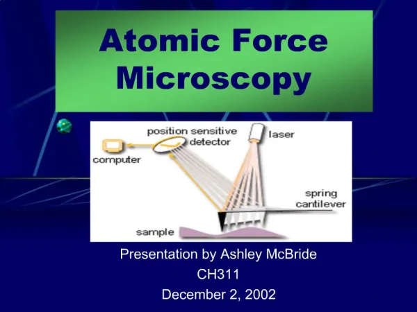
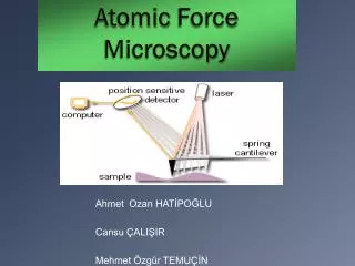
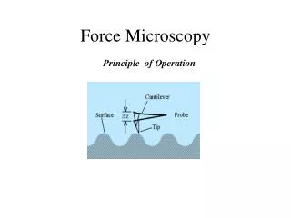
Force Microscopy. Principle of Operation. Force Microscopy. Basic Principle of Operation: detecting forces between a mass attached to a spring (cantilever), that feels some force when it is brought very close to the surface. Ideally the mass (tip) would not damage the surface.

Force Microscopy
E N D
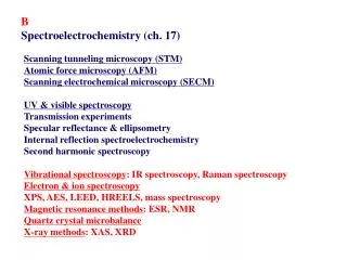
Presentation Transcript
Force Microscopy Principle of Operation
Force Microscopy • Basic Principle of Operation: detecting forces between a mass attached to a spring (cantilever), that feels some force when it is brought very close to the surface. Ideally the mass (tip) would not damage the surface. • Sensor that responds to a force and a detector that measures it. • The sensor-a cantilever beam with an effective spring constant k, moves in accordance with the forces acting on its tip
Force Microscopy • Frequency of atoms vibration, , at room temperature ~ 1015 Hz • The mass, m, of an atom ~ 10-30 kg • The effective spring constant, k, between atoms is k=2m1N/m
Materials Characterization Courtesy Dr. Z. Barkai
AFM Images2. Carbon nanotube 3. Human chromosomes TappingMode AFM image of single carbon-nanotube molecule on electrodes. These images represent an important breakthrough where we measured electronic transport through a single nanotube molecule for the first time. 530nm x 300nm scan courtesy C. Dekker and Sander Tans, Delft University of Technology, Department of Applied Physics and DIMES, The Netherlands.
Materials Characterization Courtesy Dr. Z. Barkai
AFM Images1. Au (111) High resolution scan of Au (111) surface, with reconstruction strips (inset) hexagonal atomic structure. Scan size: 5nm; inset: 20 nm
Laser Contact - Atomic Force Microscopy Fv Fc Total Repulsive Total Atractive All rights reserved @ Norbert
AFM -Cantilevers Diamond-coated AFM tip FIB Sharp Tip • Pyramidal, tetrahedral, or conical tips are the most common tip shapes Gold-coated Si3N4 Tip
AFM -Cantilevers • Depositing a Si3N4 layer on an etch pit in Si • Tips are broader then Si conical tips, harder, and thinner (stress in the film) • not suitable for non-contact (small thickness, small )
AFM -Resolution • STM-single atom interaction • AFM-several atoms on tip interact with several atoms on surface STM • In contact, not necessarily a single atom contact, radius of contact ~(Rd)1/2 (d-penetration depth, R-radius of tip) AFM
AFM -Resolution • Interaction of atom 1 : t=0 different from interaction of atom 3,2 • Each tip atom produces a signals with offset to each other • Periodicity reproduced but no true atomic resolution