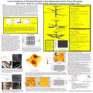
Atomic Force Microscopy & Chemical Force Microscopy
Atomic Force Microscopy & Chemical Force Microscopy. Biological systems can only be fully understood if their structure is known Structural Biology : the science investigating the structure and function of the components of living systems . Traditional methods

Atomic Force Microscopy & Chemical Force Microscopy
E N D
Presentation Transcript
Atomic Force Microscopy & Chemical Force Microscopy
Biological systems can only be fully understood if their structure is known • Structural Biology : the science investigating the structure and • function of the components of living systems. • Traditional methods • - X-ray crystallography, NMR : Too complicated, limited • size (200 Kd, 40 Kd) • - Electron microscopy • - Impossible for observation under physiological conditions
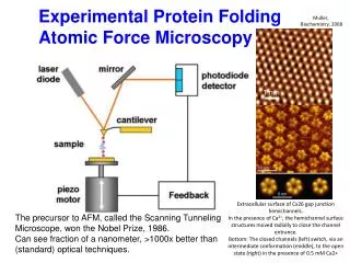
AFM : Atomic force microscope • High resolution type of scanning probe microscope • Invented by Binnig, Quate, Gerber in 1986 • To determine the surface topography of native biomolecules at sub-nanometer resolution not only under physiological conditions, but while biological processes are at work. • One of the foremost tools for imaging, measuring, and manipulating matter at the nanoscale • High signal-to-noise (S/N) ratio : details topological information • is not restricted to crystalline specimens. • Utilize a sharp probe moving over the surface of a sample in a raster scan. • The probe is a tip on the end of a cantilever which bends in response to the • force between the tip and the sample surface
Principle of AFM • Scan an object point by point using a cantilever tip • Determine the forces between the tip and the sample based on a deflection of the cantilever according to Hook’s law. • The cantilever obeys Hook’s law for small displacement, and • the interaction force between the tip and the sample can be • determined. • Measure the deflection using a laser spot reflected from the top of the cantilever into an array of photodiodes.
Schematic of AFM using the light deflection mode • As the cantilever flexes, the light from the laser is reflected onto the photo-diode • Change in the bending of the cantilever is measured • The movement of the tip or sample is performed by an extremely precise positioning device made from piezo-electric ceramics, mostly in the form of a tube scanner. • The scanner moves the sample or the cantilever in x, y, and z direction at sub-angstrom resolution
Force curve as a function of the distance between the tip and the surface : Van der waals force Van der waals force f(r) ~ -1/r6 +1/r12 10-7 ~10-11 N
Feedback operation • If the tip were scanned at a constant height, the tip would collide with the surface, causing damage • A feedback mechanism is employed to adjust the tip-to sample distance to maintain a constant force between the tip and the sample • The sample is mounted on a piezoelectric tube that can move the sample in the z-direction for maintaining a constant force, and the x and y directions for scanning the sample. • Operation in two principle modes - With feedback control: the positioning piezo responds to any changes in force that are detected, and alter the tip-sample separation to restore the force to a predetermined value constant force mode a fairly faithful topographical image - Without feedback control: Constant height or deflection mode - Useful for imaging very flat sample at high resolution - A small amount of feedback-loop gain to avoid problems with thermal drift or damaging the tip and/or cantilever.
Imaging modes • Contact mode (Static mode) • Dynamic force mode - Non-contact mode - Intermittent contact mode (Tapping mode) - Force modulation mode
Contact mode • The most common method • The tip and sample remain in close contact, namely in the repulsive regime of the inter-molecular force curve as the scanning proceeds • Repulsive force : 1~10 nN • Deflection of cantilever with a low spring constant • Determine the reflection of laser from the top of the cantilever using a photodiode • Alter the tip-sample separation to restore the force to a predetermined valuescanner • Image the surface by analyzing the changes in z-direction
Contact mode • Very sensitive to a small force • Measuring a displacement as small as 0.01nm • Image with high resolution • Damage of the sample and/or tip , cantilever • Large lateral forces on the sample as the tip is • effectively dragged across the surface • Combined effects from the capillary forces of the water contamination layer
Dynamic Modes • Distance between the tip and the sample : 2 – 30 nm • Attractive force : 0.1 ~ 0.01 nN • Vibration of cantilever around its resonance frequency • Due to a too small force, it is impossible to determine directly the deflection • of cantilever • Measure the changes in the frequency (fo) of cantilever caused by interaction • between the sample and cantilever • Oscillation of the cantilever : mechanical, magnetic or piezoelectric in air. • Oscillation in liquid is driven acoustically
Non-contact mode • The tip remains at all times in the attractive part of the interaction curve, and scans above the surface with a relatively small amplitude. • The tip may jump into contact with the surface if the attractive forces exerted are greater than the spring constant of the cantilever. • Much stiffer cantilever is required • Resonant frequency : 150 – 300 kHz • Almost unusable in liquid system as the damping of the small cantilever oscillation by water or other liquids is too large and the signal disappears. • Low resolution with a minimum value of around 1 nm
Typical Characteristics • Resolution similar to contact mode • Removal of the lateral forces • → No surface damage • Sharp cantilever with a high resonance frequency and • large spring constant (more stiff cantilever)
Dynamic modes • Resonant frequency of cantilever feff = 1/2π (keff / m)1/2 K eff : the spring constant of the cantilever, m : the mass of the cantilever • As the tip approaches the surface, the effective mass of the cantilever will change due to the attractive forces acting on the point. Accordingly, the resonant frequency of the cantilever, feff, will change. • Changes in the resonant frequency causes the variation in amplitude or the phase shift • Two modes of detection are possible : amplitude or phase shift • By defining the set point in terms of the signal amplitude or phase shift, the feedback loop is engaged.
Intermittent contact mode : Tapping mode • The next most common mode • The cantilever moves rapidly with a large oscillation between the repulsive and attractive regimes of the force curve. • The maximum forces applied to the surface may be lower or higher than those experienced in the contact mode, but such forces are not applied constantly, lowering drag forces on the sample. • Stiff cantilever with resonant frequencies in the range of 200- 400 kHz To break free of water contamination : damping problem • The problem of capillary forces is removed. • The phase shift is highly sensitive to the tip-sample interaction and generates information on the mechanical properties of the sample. • Phase shifting may occur via adhesion between the tip and the sample or by a viscoelastic response of the sample.
Force modulation mode • Combine the oscillation of the cantilever with scanning in the contact mode. • Low oscillation between 1 – 5 kHz • The information extracted concerns the mechanical and viscoelastic properties of the sample • Useful for imaging the sample containing composite materials.
Cantilever • Material : Si, Si3N4 • Stiffness • soft : contact mode (thickness : ~ 0.6㎛) • stiff : dynamic force (thickness : ~ 4㎛) • Spring Constant (k) : 0.1 ~ 10 N/m • Resonance frequency : 10~100 kHz
Artifacts related to tip size and shape • The sharpness of the scanning tip : One of the most important factors affecting the resolution • Tip convolution - Broadening :Occur when the radius of the tip curvature is comparable or greater than the size of the feature to be imaged. As the tip scans over the surface, the sides of the tip make contact before the apex, and the microscope begins to respond to the feature : Tip convolution. - Compression : The tip is over the feature - Interaction forces : Change in force interaction due to the chemical nature of the tip - Aspect ratio : when imaging steep sloped features
Tip deconvolution effects Observed width W = (8dR)1/2
Images of AFM Contact mode Dynamic force mode
Chloroplast ATP synthase is revealed to be composed of 14 subunits
AFM topographs of purple membrane from Halobacterium salinariumPurple membrane consists of 25 % lipid and 75 % bacteriorodopsin. The light driven proton pump comprises 7 transmembrane a-helices that surround the photoactive retinal
AFM images of the cytoplasmic surface of the hexagonally packedintermediate layer of the bacterium Deinoccocus radiodurans Protruding protein cores
Dip pen-nanolithography using AFM MHA : 16-mercaptohexadecanoic acid Passivated by 11-mercaptoundecyl-tri(ethylene glycol) Lee et al. Science, 295, 1702-1705 (2002)
Chemical Force Microscope Force-Distance Analysis • When the tip is placed at a fixed point on the sample and move in the vertical direction to the surface and then retracted from the surfacein place of scanning, the deflection of the cantilever can be measured as it moves. • The cantilever is in the repulsive, contact region of the cycle, and the adhesion interactions between the tip and the surface • The deflection of the cantilever will provide information on the mechanical properties of the material during the part of the approach and the retraction.
(a) and (b) When the sample is hard and incompressible, as would be seen with glass, ceramics or metallic surfaces, the tip will simply approach the surface, jump into contact and then bend ; the retraction curve will be the same. (c) For more compressible samples, the curve will be expected to resemble that shown in (c) and information on the mechanical properties of the sample may be extracted .
Adhesion force Fadh : R : size of the sphere (radius) W : work of adhesion
Work of adhesion : Dupre equation - For a typical hydrocarbon, γw= 435, γHC = 108, γHC/W = 304 J/mol/A2 - For the 2.7 nN rupture force required to separate the complementary DNA interface, we calculate 1.6 * 10-4 J/m2 for the work of adhesion.
CH3/CH3 : 1.0+0.4 nN CH3/COOH : 0.3 + 0.2 nN
Chemical force imaging : Chemical sensitive imaging • AFM probe tips are covered by particular chemical functional groups (-CH3, NH2, COOH or more exotic biological molecules) • Scanned over a sample to detect adhesion differences between the species on the tip and those on the surface of the sample • Chemical imaging of structures present on the surface due to differences in interactions between the tip and sample
Small molecule DNA binding mode • Cell replication and gene expression : specific DNA-protein interactions • Blocking of the processes by small molecules : Therapeutic agents • Binding modes of small molecules : Understanding of their functions and development of new drugs • Binding through interactions, groove binding, and covalent attachment - Cisplatin ( cis-platinum diammine dichloride) : the cross-linking anti-cancer drug - Berenil : the anti-trypanosomal minor groove binder - Ethidium bromide : the intercalating dye
Four Bases in DNA :A,G,C,T Pyrimidine (질소와 탄소로 구성된 6각형 고리 ) : thymine, cytosine Purine (질소와 탄소로 구성된 6각형과 5각형의 이중 고리) : adenine, guanine