Microbiology 155
Microbiology 155. Lecture 2- Microscopy. Microscopy. Properties of light Wavelengths of light= colors. The visible spectrum Ranges from 420-680 nm. Resolution of light

Microbiology 155
E N D
Presentation Transcript
Microbiology 155 Lecture 2- Microscopy
Microscopy • Properties of light • Wavelengths of light= colors. The visible spectrum • Ranges from 420-680 nm Resolution of light The resolution which is the ability to see items as separate and distinct entities is determined by the wavelength of light used in the microscope
Resolution and Images • For two points to be seen as separate the light has to be able to pass between them • If the points are very close and the light is not able to pass between, the image will appear as fuzzy. • The light microscope used in class is unable to resolve two points that are closer together than 220 nm • The average wavelength of light is 550 nm
The Resolving Power of a Lens • The resolving power of a lens is a numerical measure of the resolution that can be attained with that lens. • The smaller the distance between two points that can be resolved the stronger the power of the lens • Ultra violet light has a wavelength of 100- 400 nm, As a result It can resolve points that are 110 nm apart.
The Numerical Aperture ( NA) • NA= the numerical apertures . The numerical aperture is a measure of the light that is collected and directed through the microscope oculars. The part of the microscope that collects the light is referred to as the condenser. • RP= Lambda/2NA
Reflection • If light strikes an object and bounces back- reflection has occurred • Refraction • If light bends as it passes from one medium to another - through different densities- it is bent or refracted • The index of refraction for a material is the measure of the speed at which the light passes through the material.
Bright Field Microscopy ( used to visualize stained slides of bacteria
Dark Field A microscope adapted for dark field has a condenser that prevents light from being transmitted through the specimen on the slide. The background appears dark and the bacterium or organism appears to glow Used to show contrast such as with spirochetes.
Phase Contrast Microscopy This microscopic technique is used to view live cells. It amplifies differences in the cellular structure and contents. These are called refractive differences .
NOMARSKI DIFFERENTIAL INTERFERENCE • Operate like phase contrast microscopes but with much greater resolution • This produces almost a three dimensional image
Fluorescence Microscopy Fluorescent stains use colored molecules that become excited when short wave lengths of light shine on them These colored molecules are called fluorochromes
Fluorescence- Antibody Staining • Antibodies are molecules produced by the immune system in response to invaders such as bacteria, protozoans, and viruses. • Antigens are foreign substances that are present • An antigen- antibody reaction is a specific reaction • This can be linked to the fluorescent staining
Electron Microscopy • Use electron beam as light source • Electrons have shorter wavelength than visible light • Electromagnetic lenses help to magnify and focus • Resolution improves to .2um • Photographs can be taken although objects can not be directly visualized these are known as • Electron micrographs
Transmission Electron Microscopy TEM • The electron beam is concentrated and passed through an object • The object is stained with heavy metal atoms • The interaction of the electrons with the object produces an image • It is a flat image
Transmission Electron Microscopy showing the internal structure. These cells are dividing by the process of fission
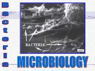
Scanning Electron Microscopy- SEM • In scanning electron microscopy the cells are stained on the outside. The electron beams are shot at the cells and bounce back. • They are caught by a recording device that transmits the electrons to a TV screen • The image can be visualized on the screen
Electron Micrograph of Blood Cells These cells are reacting to the presence of bacteria in the blood stream.
Freeze Etching and Freeze Fracture Cells are frozen in liquid nitrogen The cells are broken open or fractured by touching with a knife The inner membranes and structures of the cell are revealed “ moonscape” In freezing etching - the surface of the “moonscape” is layered with a heavy metal- The surface is then viewed with an electron microscope
Stains • Stains have special characteristics that allow them to bind to the cell wall of bacteria • Bacterial cell walls are negative. • Bacterial stains are positively charged • Stains that are positively charged are cationic
Stain terminology • Simple stain- contains one dye molecule • Differential stains are composed of two or more dyes • Negative stains color the background • Flagellar stains add layers of dye or metal to the surface of flagella
Visualization of Bacterial Cells Staining Protocols Stain components of cell walls Gram - These bacteria stain pink with the gram stain Gram + These bacteria stain purple with the gram stain
Gram Stain Protocol • Smear slide with cells from culture of bacteria • Heat fix. Move slide through the flame • Cover smear with Crystal Violet( Purple Stain)- 1 min • Rinse with water. Allow water to rinse purple stain off the slide • Cover the slide with Iodine. Iodine is a mordant. It helps cells sticks to slide
Gram Stain ( continued) • Place a few drops of alcohol on the slide • This will remove any stain that is not permanently attached to the cell walls • Place drops of safranin over smear • Rinse and air dry
Acid Fast Staining- specialized technique Acid fast bacteria retain carbol fuschin and appear red Non- acid fast accept the methylene blue counterstain and appear blue Mycobacterium is identified with this staining procedure Leprosy and tuberculosis
Acid fast staining- arrows pointing to acid fast bacteria( reddish)
Spore staining- important to bacteria called spore formers clostridium,botulism, tetanus, and anthrax Endospores retain malachite green stain Vegetative cells accept safranin and appear red
Schaeffer-Fulton spore stain- makes spores easier to visualize