Download
1 / 50
510 likes | 549 Vues
Explore the brain's principal parts, blood supply, protective coverings, blood-brain barrier, and cerebrospinal fluid. Learn about the medulla oblongata, pons, midbrain, and reticular formation. Discover the vital role of the cerebellum in muscle coordination and equilibrium maintenance.

E N D
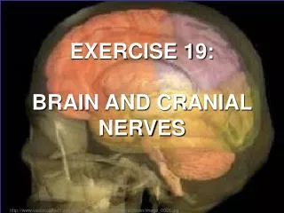
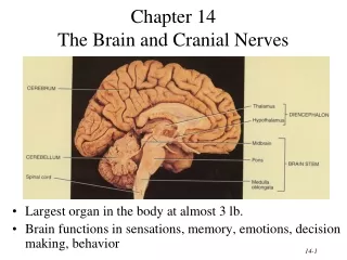
Chapter 14The Brain and Cranial Nerves • Largest organ in the body at almost 3 lb. • Brain functions in sensations, memory, emotions, decision making, behavior
Principal Parts of the Brain • Cerebrum • Diencephalon • thalamus & hypothalamus • Cerebellum • Brainstem • medulla, pons & midbrain
Blood Supply to Brain • Arterial blood supply is branches from circle of Willis on base of brain (page 699 or next slide) • Vessels on surface of brain----penetrate tissue • Uses 20% of our bodies oxygen & glucose needs • blood flow to an area increases with activity in that area • deprivation of O2 for 4 min does permanent injury • at that time, lysosome release enzymes
Venous Drainage • Superior saggital sinus etc- in subdural
Protective Coverings of the Brain • Bone, meninges & fluid • Meninges same as around the spinal cord • dura mater • arachnoid mater • pia mater • Dura mater extensions • falx cerebri • tentorium cerebelli • falx cerebelli
Blood Brain Barrier • Blood-brain barrier (BBB) • protects cells from some toxins and pathogens • proteins & antibiotics can not pass but alcohol & anesthetics do • tight junctions seal together epithelial cells, continuous basement membrane, astrocyte processes covering capillaries
What goes in and What Doesn’t • Lipid Soluble- O2, CO2, ETOH, anesth all pass easily the BBB • Watersoluble, glucose, urea, creatinine, ions pass slowly or in the case of glucose, via active transport only • BBB is not uniform throughout, some areas are more permeable and thus bacteria can get in
Cerebrospinal Fluid (CSF) • 80-150 ml (3-5oz) • Clear liquid containing glucose, proteins, & ions • Functions • mechanical protection • floats brain & softens impact with bony walls • chemical protection • optimal ionic concentrations for action potentials • circulation • nutrients and waste products to and from bloodstream
Origin of CSF • Choroid plexus = capillaries covered by ependymal cells • 2 lateral ventricles, one within each cerebral hemisphere • roof of 3rd ventricle • fourth ventricle
Drainage of CSF from Ventricles • One median aperture & two lateral apertures allow CSF to exit from the interior of the brain
Cerebral Perfusion Pressure • CPP = MABP – ICP • Cerebral auto regulation
Hydrocephalus • Blockage of drainage of CSF (tumor, inflammation, developmental malformation, meningitis, hemorrhage or injury • Continued production cause an increase in pressure --- hydrocephalus • In newborn or fetus, the fontanels allow this internal pressure to cause expansion of the skull and damage to the brain tissue • Neurosurgeon implants a drain shunting the CSF to the veins of the neck or the abdomen
Medulla Oblongata • Continuation of spinal cord • Ascending sensory tracts • Descending motor tracts • Nuclei of 5 cranial nerves • Cardiovascular center • force & rate of heart beat • diameter of blood vessels • Respiratory center • medullary rhythmicity area sets basic rhythm of breathing • Information in & out of cerebellum • Reflex centers for coughing, sneezing, swallowing etc
Injury to the Medulla • Hard blow to the back of the head may be fatal • Cranial nerve malfunctions on same side as injury;loss of sensation or paralysis of throat or tongue; irregularities in breathing and heart rhythm
Pons • One inch long • White fiber tracts ascend and descend • Pneumotaxic & apneustic areas help control breathing • Middle cerebellar peduncles carry sensory info to the cerebellum • Cranial nerves 5 thru 7
Midbrain • One inch in length • Extends from pons to diencephalon • Cerebral aqueduct connects 3rd ventricle above to 4th ventricle below • Visual reflex centers and auditory relay (startle reflex)
Midbrain in Section • Cerebral peduncles---clusters of motor & sensory fibers • Substantia nigra---helps controls subconscious muscle activity • Red nucleus-- rich blood supply & iron-containing pigment • cortex & cerebellum coordinate muscular movements by sending information here from the cortex and cerebellum
Reticular Formation • Scattered nuclei in medulla, pons & midbrain • Reticular activating system • alerts cerebral cortex to sensory signals (sound of alarm, flash light, smoke or intruder) to awaken from sleep • maintains consciousness & helps keep you awake with stimuli from ears, eyes, skin and muscles • Motor function is involvement with maintaining muscle tone
Cerebellum • 2 cerebellar hemispheres and vermis (central area) • Function • correct voluntary muscle contraction and posture based on sensory data from body about actual movements • sense of equilibrium
Cerebellum • Transverse fissure between cerebellum & cerebrum • Cerebellar cortex (folia) & central nuclei are grey matter • Arbor vitae = tree of life = white matter
Diencephalon Surrounds 3rd Ventricle • Surrounds 3rd ventricle • Superior part of walls is thalamus • Inferior part of walls & floor is hypothalamus
Thalamus • 1 inch long mass of gray mater in each half of brain (connected across the 3rd ventricle by intermediate mass) • Relay station for sensory information on way to cortex • Crude perception of some sensations
Thalamic Nuclei • Nuclei have different roles • relays auditory and visual impulses, taste and somatic sensations • receives impulses from cerebellum or basal ganglia • anterior nucleus concerned with emotions, memory and acquisition of knowledge (cognition)
Hypothalamus • Dozen or so nuclei in 4 major regions • mammillary bodies are relay station for olfactory reflexes;infundibulum suspends the pituitary gland • Major regulator of homeostasis • receives somatic and visceral input, taste, smell & hearing information; monitors osmotic pressure, temperature of blood
Functions of Hypothalamus • Controls and integrates activities of the ANS which regulates smooth, cardiac muscle and glands • Synthesizes regulatory hormones that control the anterior pituitary • Contains cell bodies of axons that end in posterior pituitary where they secrete hormones • Regulates rage, aggression, pain, pleasure & arousal • Feeding, thirst & satiety centers • Controls body temperature • Regulates daily patterns of sleep
Cerebrum (Cerebral Hemispheres) • Cerebral cortex is gray matteroverlying white matter • 2-4 mm thick containing billionsof cells • grew so quickly formed folds(gyri) and grooves (sulci or fissures) • Longitudinal fissure separates left & right cerebral hemispheres • Corpus callosum is band of white matter connecting left and right cerebral hemispheres • Each hemisphere is subdivided into 4 lobes
Longitudinal fissure (green) Frontal lobe Central sulcus (yellow) precentral & postcentral gyrus Parietal lobe Parieto-occipital sulcus Occipital lobe Lateral sulcus (blue) Temporal lobe Lobes and Fissures
Basal Ganglia • Connections to red nucleus, substantia nigra & subthalamus • Input & output with cerebral cortex, thalamus & hypothalamus • Control large automatic movements of skeletal muscles
Limbic System • Parahippocampal & cingulate gyri & hippocampus • Emotional brain--intense pleasure & intense pain • Strong emotions increase efficiency of memory
Brain Injuries • Causes of damage • displacement or distortion of tissue at impact • increased intracranial pressure • infections • free radical damage after ischemia • Concussion---temporary loss of consciousness • headache, drowsiness, confusion, lack of concentration • Contusion--bruising of brain (less than 5 min unconsciousness but blood in CSF) • Laceration--tearing of brain (fracture or bullet) • increased intracranial pressure from hematoma
Types of Injuries Cont • Epidural bleed –arterial- Middle meningeal artery – retrograde unconsiousness -treated Burr holes • SubDural Bleed –venous- bridging veins • Subarchnoid bleed- arterial-Circle of Willis • Moderate axonal injury • Diffuse axonal tearing injury
Sensory Areas of Cerebral Cortex Receive sensory information from the thalamus Primary somatosensory area = postcentral gyrus = 1,2,3 Primary visual area = 17 Primary auditory area = 41 & 42 Primary gustatory area = 43
Motor Areas of Cerebral Cortex • Voluntary motor initiation • Primary motor area = 4 = precentral gyrus • controls voluntary contractions of skeletal muscles on other side • Motor speech area = 44 = Broca’s area • production of speech -- control of tongue & airway
Association Areas of Cerebral Cortex (FYI) • Somatosensory area = 5 & 7 (integrate & interpret) • Visual association area = 18 & 19 (recognize & evaluate) • Auditory association area(Wernicke’s) = 22(words become speech) • Gnostic area = 5,7,39 & 40 (integrate all senses & respond) • Premotor area = 6 (learned skilled movements such as typing) • Frontal eye field =8 (scanning eye movements such as phone book)
Aphasia • Language areas are located in the left cerebral hemisphere of most people • Inability to use or comprehend words = aphasia • nonfluent aphasia = inability to properly form words • know what want to say but can not speak • damage to Broca’s speech area • fluent aphasia = faulty understanding of spoken or written words • faulty understanding of spoken or written words • word deafness = an inability to understand spoken words • word blindness = an inability to understand written words • damage to common integrative area or auditory association area
Hemispheric Lateralization • Functional specialization of each hemisphere more pronounced in men • Females have larger connections between 2 sides • Damage to left side produces aphasia • Damage to same area on right side produces speech with little emotional inflection
The Spinal Cord • Extends from the foramen magnum of the skull to L¹ or L² ; the dural and arachnoid membranes extend to the level of S², well beyond the end of the spinal cord which makes this an ideal location for a lumbar tap • 31 pairs of spinal nerves attach to the cord by paired roots and exit from the vertebral column via the intervertebral foramina to travel to the body regions they serve
The Spinal Cord • It is about the width of a thumb for most of its length, but is enlarged in the cervical and lumbosacral regions, where the nerves serving the upper and lower limbs arise • The collection of nerve roots at the inferior end of the vertebral canal is named the cauda equina
The Spinal Cord: Gray Matter and Spinal Roots • Looks like the letter “H” or a butterfly with the cross-bar of gray matter called the gray commissure that encloses the central canal
The Spinal Cord: Gray Matter and Spinal Roots • Dorsal and Ventral Roots: very short and fuse laterally to form the spinal nerves • The spinal nerves form the peripheral nervous system
The Spinal Cord: White Matter • Composed of myelinated and unmyelinated nerve fibers that allow communication between different parts of the spinal cord and between the cord and brain • (1) Ascending: up to higher centers (sensory inputs) (2) Descending: down to the cord from the brain or within the cord to lower levels (motor outputs) (3) Transversely: across from one side of the cord to the other (commissural fibers) • Ascending and descending make up most of the white matter
Spinal cord Injuries The following list outlines which muscle functions may be retained at progressively lower levels of spinal cord transection. C1–C3: no function maintained from the neck down; ventilator needed for breathing C4–C5: diaphragm, which allows breathing C6–C7: some arm and chest muscles, which allows feeding, some dressing, and propelling wheelchair T1–T3: intact arm function T4–T9: control of trunk above the umbilicus T10–L1: most thigh muscles, which allows walking with long leg braces L1–L2: most leg muscles, which allows walking with short leg braces