Fig, 47.12 (3)
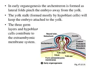
In early organogenesis the archentreron is formed as lateral folds pinch the embryo away from the yolk. The yolk stalk (formed mostly by hypoblast cells) will keep the embryo attached to the yolk. The three germ layers and hypoblast cells contribute to the extraembyonic membrane system.

Fig, 47.12 (3)
E N D
Presentation Transcript
In early organogenesis the archentreron is formed as lateral folds pinch the embryo away from the yolk. • The yolk stalk (formed mostly by hypoblast cells) will keep the embryo attached to the yolk. • The three germlayers and hypoblastcells contribute tothe extraembyonicmembrane system. Fig, 47.12 (3)
Extraembryonic membranes Cells of the yolk sac digest yolk providing nutrients to the embryo. The amnion encloses the embryo in a fluid-filled amniotic sac which protects the embryo from drying out. The chorion cushions the embryo against mechanical shocks. The allantois functions as a disposal sac for uric acid.
Mammalian Extraembryonic membranes The trophoblast gives rise to the chorion, which continues to expand into the endometrium and the epiblast begins to form the amnion.
Mammalian embryonic membranes homologous with those of shelled eggs. Chorion: completely surrounds the embryo and other embryonic membranes. Amnion: encloses the embryo in a fluid-filled amniotic cavity. Yolk sac:Develops from the hypoblast. Site of early formation of blood cells and differentiation of PGCs Allantois: develops as an outpocketing of the embryo’s rudimentary gut. Incorporated into the umbilical cord, where it forms blood vessels.