Download
1 / 8
80 likes | 181 Vues
A comprehensive guide for using the Zeiss LSM510 Confocal Microscope, including startup procedures, laser safety precautions, setting configurations, and visual setup instructions. Contact Rachel Gregory or Liz Brooks for assistance.

E N D
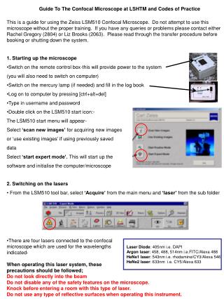
Guide To The Confocal Microscope at LSHTM and Codes of Practice • This is a guide for using the Zeiss LSM510 Confocal Microscope. Do not attempt to use this microscope without the proper training. If you have any queries or problems please contact either Rachel Gregory (2804) or Liz Brooks (2063). Please read through the transfer procedure before booking or shutting down the system. • 1. Starting up the microscope • Switch on the remote control box-this will provide power to the system • (you will also need to switch on computer) • Switch on the mercury lamp (if needed) and fill in the log book • Log on to computer by pressing [ctrl+alt+del] • Type in username and password • Double click on the LSM510 start icon:- • The LSM510 start menu will appear- • Select ‘scan new images’ for acquiring new images • or ‘use existing images’ if using previously saved • data • Select ‘start expert mode’. This will start up the • software and initialise the computer/microscope • 2. Switching on the lasers • From the LSM510 tool bar, select ‘Acquire’ from the main menu and ‘laser’ from the sub folder • There are four lasers connected to the confocal • microscope which are used for the wavelengths • indicated- • When operating this laser system, these • precautions should be followed; • Do not look directly into the beam • Do not disable any of the safety features on the microscope. • Knock before entering a room with this type of laser. • Do not use any type of reflective surfaces when operating this instrument. Laser Diode: 405nm i.e. DAPI Argonlaser: 458, 488, 514nm i.e.FITC/Alexa 488 HeNe1laser: 543nm i.e. rhodamine/CY3/Alexa 546 HeNe2laser: 633nm i.e. CY5/Alexa 633
To turn the Argon laser on, first highlight the laser name in the control window and click the ‘stand-by’ button. When the laser has warmed up a ‘Ready’ message will appear, and the laser may now be turned on, select ‘on’. The laser output (%) will need to be adjusted to create a tube current of 6.1A (see diagram). Laser output is usually adjusted to around 55%. • The HeNe and laser diode lasers can be turned on directly by first highlighting the laser names individually and then clicking the ‘on’ buttons. There’s no need to adjust the laser output (%). • 3. Set Track/Laser configurations • Select ‘config’ from the tool bar subfolder • Choose the required laser path settings from the pre-programmed configurations- • If using a single fluorophore, click on • ‘single track’. If using more than one • fluorophore click on ‘multi track’ • Click ‘config’ from the configuration • control window • Select required configuration from drop-down • menu and click ‘apply’. 6.1A
4. Setting up the microscope visually • You can toggle between scanning mode and visual mode using • the LSM toolbar • From LSM510 toolbar select ‘Micro’ • This window allows you to:- • Switch on/off transmitted light and adjust • its brightness • Select the objective to be used • Switch on/off reflected light • Select the filter to be used • Select the objective you require to image your sample. If the objective needs it, add a drop of oil onto your slide. • Place the slide ‘upside down’ and clamp into place. • It is normally best to focus using transmitted light in case the fluorescence labelling has been unsuccessful. • Switch on the transmitted light by selecting the ‘transmitted light’ in the microscope control menu. Light brightness is usually set to 5-6, (but you can go brighter if you wish). Once you have focused on your sample you can check for fluorescence. • Switch off the transmitted light and select ‘reflected light’. Choose the appropriate filter for your sample from the ‘reflector’ drop down menu. • N.B. you cannot visualise far red (i.e. Alexa 633 or CY5) with the filters, these can only be visualised by scanning.
5. Set Scanning Configurations • From the LSM510 Toolbar subfolder, select ‘Scan’ • From the Scan control box select ‘Mode’ • Standard settings:- • Frame size: 512x512 • Scan speed: 8-9 • Mode: Line • Method: Mean • Number: 1* • (*indicates the number of scans averaged to create • The final image. Increasing the number can reduce • Noise and increase quality of image, but may also • bleach the sample! Best to have 1 when optimising • scanning, and Increase to 4-8 when taking final • scan.) • From the Scan control box select ‘channels’ • The pinhole for each channel used must be altered • so they are as similar to each other as possible:- • Select the channel with the longest wavelength and • set pinhole to 1.00 Airy Unit • Select the other channels being used individually • and alter the pinhole setting so pinhole and optical • slice are as close to the settings for the longest • wavelength channel as possible • [small adjustments can be made by holding down • Ctrl] (If your sample is weak you can use >1.00 Airy • Units,but your image will no longer be conical). • Set Amp gain to 1.
6. Scanning an Image • Once you have located a field you wish to image, • return to LSM mode on the toolbar • From the Scan control box (channels) select, • ‘new’ this will open a new viewing window in • the right-hand screen • Select ‘find’. This will adjust the detector gain and • offset for your sample. • To further optimise image, select ‘palette’ from the • viewing window, then ‘range indicator’. • Select ‘fast XY’ • Select one channel and adjust the ‘detector gain’ • and ‘amplifier offset’ accordingly • When gain and offset have been optimised, stop • scanning by selecting ‘stop’. • Repeat for all channels being used (they must be • done separately). • Turn off range indicator by selecting ‘no palette’ in • palette window. • For final image increase mode number to 4-8 (see section 4) and select ‘single’ from the scan control window, under channels. You should now save this image to your directory.
7. Acquiring Phase/light images • This method allows you to image the transmitted light field. If you wish to take a DIC image • please follow the appropriate protocol. • In the configuration control window, check the ‘ChD’ • selection box towards the bottom of the window. • This will add the light channel to which ever channel • is selected at the time. It is usually advised that you use • the channel with the longest wavelength in use for this • process. • It can be removed by un-checking the box. • The offset and gain of the light channel of the light • image can be adjusted as per fluorescence. • 8. Taking Z-Series • To image a Z-series, first select ‘Z settings’ in • the scan control window. • Using the focus wheel on the microscope, alter • the focus so you are at the top of your sample, • Select ‘mark first’. • Then alter the focus so you are at the bottom • of your sample, select ‘mark last’. • By selecting Z slice, you can instruct the • software to calculate the number of slices • from this section needed to provide the optimal • interval. • You can alter the number of slices imaged • manually in the Z settings window. • When you have finished the set up, select • ‘start’.
9. Creating a database/Saving your images • To open a pre-existing database, select ‘File’ from the LSM toolbar, then ‘Open’ from the sub folder. Navigate to your database. • To create a new database, select ‘New’ from the subfolder • User databases should be saved on the D: drive, enter your database name and create a destination folder. • When you have an image you want to save, select ‘Save as’. Assign your image a name, click ‘OK’ . • N.B. It is advisable to save a successfully acquired image immediately, as they can be easily lost when scanning a new area. • If you wish to re-use offset and gain settings from a previous image, these can be • set by opening the image and selecting ‘reuse’ from the viewing window of that • image.
10. Shut down procedure • Remove your sample and clean oil from all the lenses used. • Ensure the objectives and reflective filters are left in ‘empty’ position. • If you have been contacted by the next user to leave the microscope on, do so. If you have not been contacted (please check your emails) then continue with the rest of the shut down procedure. • Switch off the lasers:- • in the laser control window, select lasers individually • HeNe1 and HeNe2 lasers may be switched off directly-select ‘off’. • The Argon laser must be allowed to cool down-select ‘standby’,then select ‘off’.The status will show ‘cooling’. The status will then change to ‘connected’ (this will take up to 5mins)the remote control must not be switched off before this time. • Switch off the mercury lamp, and fill in the log book • Exit from LSM510 and shut down the computer. • When the computer has fully shutdown, switch off the remote control • Re-cover the microscope with blue dust cover and plastic sheets • Dispose of all used tissues and other general mess in the autoclave • tin provided. • N.B. This tin is not for glass coverslips! • Ensure room is left locked if there are no other users present and return key to the Log book.