Understanding the Cell Cycle: Key Stages, Control Mechanisms, and Cancer Implications
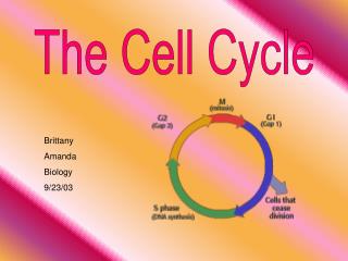
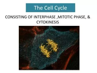
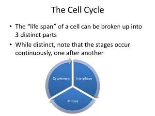
The cell cycle consists of three major stages: Interphase, Mitosis, and Cytokinesis. During Interphase, the cell undergoes G1, S, and G2 phases, where it performs basic functions, replicates DNA, and prepares for division. Mitosis follows, involving phases such as Prophase, Metaphase, Anaphase, and Telophase, ensuring equal distribution of genetic material. Cytokinesis then divides the cytoplasm, differing in animal and plant cells. Control mechanisms such as checkpoints and apoptosis regulate the cycle, while cancer arises from disruptions in this regulation.

Understanding the Cell Cycle: Key Stages, Control Mechanisms, and Cancer Implications
E N D
Presentation Transcript
The Cell Cycle Events that occur in the life of a cell. Includes 3 major stages: • Interphase • Mitosis • Cytokinesis
1. Interphase (Cell is not dividing) • G1 Phase – carries out basic functions & performs specialized activities. • duration is extremely variable • contains restriction checkpoint ~ cell “decides” to: • divide • enter a quiescent phase (G0) • die
1. Interphase (Cell is not dividing) • G0 Phase – cell maintains specialized characteristics, but does not divide Ex. neurons & muscle cells
1. Interphase (Cell is not dividing) • S Phase – cell replicates chromosomes & synthesizes proteins • animal cells replicate centrioles as well
1. Interphase (Cell is not dividing) • G2 Phase - cell synthesizes additional proteins (ex. tubulin) & assembles/stores membrane material
Mitosis (M phase) – Equal distribution of replicated genetic material. • Five steps: • Prophase • Prometaphase • Metaphase • Anaphase • Telophase
Mitosis – Prophase • replicated chromosomes condense • centrosomes separate & migrate toward opposite sides of cell • mitotic spindle forms (microtubules grow out from centrosomes) • nucleolus disappears
Mitosis – Prometaphase • nuclear membrane breaks down • spindle fibers attach to centromeres of chromosomes
Mitosis – Metaphase • chromosomes are lined up single-file along equator of mitotic spindle
Mitosis – Anaphase • Centromeres part, sister chromatids (now called chromosomes) separate • chromosomes move toward opposite poles
Mitosis – Telophase • mitotic spindle breaks down • chromosomes decondense • nuclear membranes reform around two nuclei • nucleoli reappear
Cytokinesis • Distribution of cytoplasm to daughter cells • begins during anaphase or telophase • differs in animal & plant cells
Cytokinesis in animal cells • Cleavagefurrow (slight indentation) forms around equator of cell • Actin & myosin microfilaments act like a drawstring to pinch the cell in two • Usually an equal division
Cytokinesis in plant cells • phragmoplast (microtubule structure) forms in cytoplasm & traps vesicles containing cell wall material • vesicles fuse, forming a cell plate across midline of cell • cell plate gives rise to two primary cell walls
Does cytokinesis always accompany karyokinesis? Karyokinesis in the absence of cytokinesis results in a syncytium(mass of multinucleated cells).
Control of the Cell Cycle Checkpoints - groups of interacting proteins that ensure cell cycle events occur in the correct sequence.
Shortening of telomeres - loss of telomere DNA signals cell to stop dividing. Some cells produce telomerase(enzyme that continually adds telomere DNA).
Contact Inhibition - healthy cells stop dividing when they come in contact with other cells.
Hormones - stimulate cell division. Ex. Estrogen stimulates uterine cell division Growth factors - proteins that stimulate local cell division. Ex. Epidermal growth factor (EGF) stimulates epithelial cell division filling in new skin underneath a scab Interaction of kinases & cyclins - activate genes that stimulate cell division.
B. Apoptosis Programmed cell death; part of normal development.
C. Cancer (loss of cell cycle control) Condition resulting from excess cell division or deficient apoptosis. Characteristics of Cancer Cells: • can divide uncontrollably & eternally • are heritable & transplantable • lack contact inhibition • readily metastasize • exhibit angiogenesis • exhibit genetic mutability
Causes of Cancer: • Over-expression of oncogenes Oncogenes are genes that trigger limited cell division. • Inactivation of tumor suppressor genes Tumor suppressor genes prevent a cell from dividing or promote apoptosis.
Normal functioning of oncogenes & tumor suppressor genes may be affected by environmental factors: • carcinogens • radiation • viruses • diet • exercise habits
Meiosis - formation of gametes • Somatic cells – body cells • In contrast to mitosis (occurs in somatic cells), gametes (eggs or sperm) are produced only in gonads (ovaries or testes). • In the gonads, cells undergo a variation of cell division (meiosis) which yields four daughter cells, each with half the chromosomes of the parent. • In humans, meiosis reduces the number of chromosomes from 46 to 23 • Chromosomes #1 through 22 – autosomal • Chromosome #23 – sex
Meiosis - formation of gametes • Fertilization fuses two gametes together and doubles the number of chromosomes to 46 again. • Organisms inherit single copy of each gene from each parent • These copies are segregated from each other during formation of the gametes • Homologous – corresponding male and female chromosomes