Visualization of Salmonella Utilizing GFP Overlays and Actin Interactions in Infected Cells
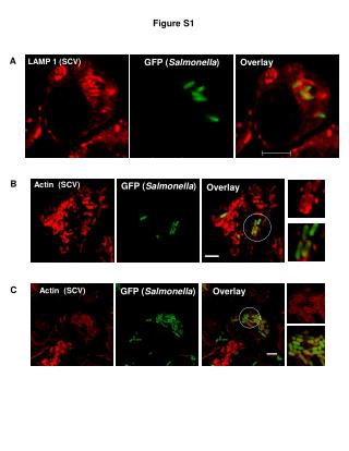
This series of figures showcases the use of GFP labeling to visualize Salmonella in infected cells. Figure S1 illustrates the overlay of GFP and Actin in SCV, highlighting the localization and interaction of Salmonella within host cells. Subsequently, Figures S2-S8 present various experimental setups, including SCV membrane examination, Nucleus interactions, and infection analyses at different multiplicities of infection (MOI). The results detail the dynamics of Salmonella in uninfected and infected conditions, providing insights into bacterial behavior and cellular response.

Visualization of Salmonella Utilizing GFP Overlays and Actin Interactions in Infected Cells
E N D
Presentation Transcript
Figure S1 A GFP (Salmonella) Overlay LAMP 1 (SCV) B Actin (SCV) GFP (Salmonella) Overlay C GFP (Salmonella) Overlay Actin (SCV)
Figure S2 A * Nucleus * * * * * * B * * * * * * * C SCV membrane Plane of section observed in the images shown above Salmonella
Figure S4 A B LAMP 1 LAMP 1 and Salmonella * * * * * * * * * *
Figure S5 Drp-1 Salmonella Overlay Drp-1 Drp-1 Salmonella Overlay Salmonella (LPS)
S.aureus Uninfected S. Typhimurium Figure S6 157 ± 3 23.3 ± 2.6*** 120.7 ± 5.5
Figure S7 Uninfected DssaV infected WT infected
Figure S8 A Uninfected Infected (MOI-50) B Uninfected Infected (MOI-50)