Download
1 / 34
370 likes | 593 Vues
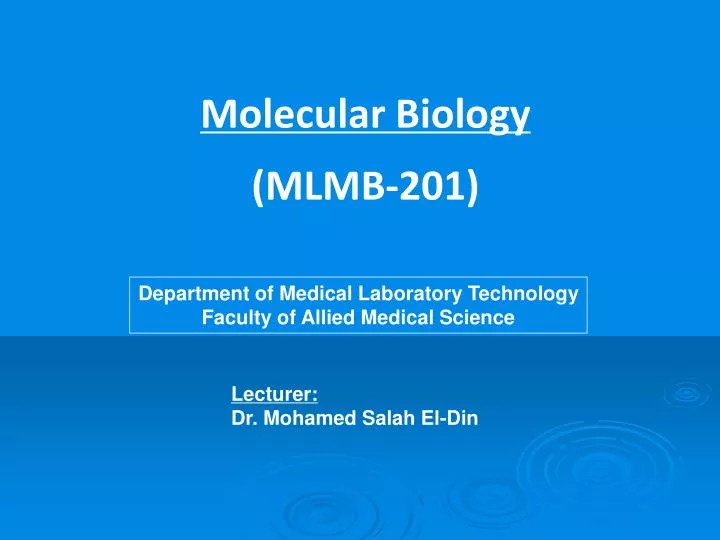
Molecular Biology (MLMB-201). Department of Medical Laboratory Technology Faculty of Allied Medical Science. Lecturer: Dr. Mohamed Salah El-Din. Intended Learning Outcomes (ILO’s): Molecular biology course provides an overview of the molecular basis to cell structure and function.

E N D
Molecular Biology (MLMB-201) Department of Medical Laboratory Technology Faculty of Allied Medical Science Lecturer: Dr. Mohamed Salah El-Din
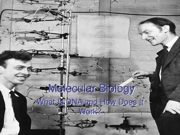
Intended Learning Outcomes (ILO’s): • Molecular biologycourse provides an overview of the molecular basis to cell structure and function. • This course focuses on the structure, biosynthesis and function of DNA and RNA on the molecular level and how these interact among themselves and with proteins. Molecular biology techniques are essential for modern biological and medical research. This course will give you an introduction to DNA and RNA standard techniques. • Student will have basic knowledge of: • Cell organization. • DNA structure and function. • DNA Extraction. • RNA structure and function. • RNA Extraction. • Gene expression and protein biosynthesis. • Agarose gel electrophoresis for DNA/RNA; and SDS-PAGE for protein. • Polymerase Chain Reaction (PCR) – Theory, Types, Application. • Gene library and screening • DNA sequencing
STRUCTURE AND FUNCTION OF RNA • RNA is structurally similar to DNA! • Both nucleic acids are sugar-phosphate polymers • Both have nitrogen bases attached to the sugars of the backbone
But there are several importantdifferences! They differ in composition: The sugar in RNA is ribose, not the deoxyribose in DNA. The base uracil is present in RNA instead of thymine. They also differ in size and structure: RNA molecules are smaller (shorter) than DNA molecules, RNA is single-stranded, not double-stranded like DNA. Another difference between RNA and DNA is in function: 1. DNA has only one function-STORING GENETIC INFORMATION in its sequence of nucleotide bases. 2. But there are three main kinds of ribonucleic acid (each of which has a specific job to do.)
The RNA polymerases • The RNA polymerases are huge multi-subunit protein complexes. • Three kinds are found in eukaryotes: • RNA polymerase I (Pol I). • It transcribes therRNA genes for the precursor of the 28S, 18S, and 5.8S • molecules (and is the busiest of the RNA polymerases). • RNA polymerase II (Pol II; also known as RNAP II). • It transcribes protein-encoding genes into mRNA (and also the snRNA genes). • RNA polymerase III (Pol III). • It transcribes the 5S rRNA genes and all the tRNA genes.
Initiation of Transcription by RNA Polymerase • Legend: • In order to begin transcription, RNA polymerase requires a number • of general transcription factors (called TFIIA, TFIIB, and so on). • The promoter contains a DNA sequence called the TATA box, • which is located 25 nucleotides away from the site where transcription is initiated. • The TATA box is recognized and bound by transcription factor TFIID, which then enables the adjacent binding of TFIIB (C). • (D) The rest of the general transcription factors as well as the RNA • polymerase itself assemble at the promoter. • (E) TFIIH then uses ATP to phosphorylate RNA polymerase II, • changing its conformation so that the polymerase is released • from the complex and is able to start transcribing. As shown, • the site of phosphorylation is a long polypeptide tail that extends from the polymerase molecule.
The steps: • Synthesis of the cap. This is a modified guanine (G) which is attached to the 5′ end • of the pre-mRNA as it emerges from RNA polymerase II (RNAP II). The cap protects • the RNA from being degraded by enzymes that degrade RNA from the 5′ end. • Step-by-step removal of introns present in the pre-mRNA and splicing of the • remaining exons. This step is required because most eukaryotic genes are split. • It takes place as the pre-mRNA continues to emerge from RNAP II. • Synthesis of the poly(A) tail. This is a stretch of adenine (A) nucleotides. • When a special poly(A) attachment site in the pre-mRNA emerges from RNAP II, • the transcript is cut there, and the poly(A) tail is attached to the exposed 3′ end. • This completes the mRNA molecule, which is now ready for export to the cytosol. • (The remainder of the transcript is degraded, and the RNA polymerase leaves the DNA.)
Main Kinds of RNA • Ribosomal RNAs-exist outside the nucleus in the cytoplasm • of a cell in structures called ribosomes. Ribosomes are small, • granular structures where protein synthesis takes place. • Each ribosome is a complex consisting of about 60% ribosomal RNA • (rRNA) and 40% protein. 2. Messenger RNAs-are the nucleic acids that "record“ information from DNA in the cell nucleus and carry it to the ribosomes and are known as messenger RNAs (mRNA). 3. Transfer RNAs-The function of transfer RNAs (tRNA) is to deliver amino acids one by one to protein chains growing at ribosomes
Ribosomal RNA • The ribosome is a large machinery (~ 20 nm in diameter, 70S sedimentation rate for bacterial ribosomes) • and is made of two subunits: • large subunit (~50 S) • is made of two ribosomal RNA (5S and 23S) and several ~34 proteins • 2. small subunit (~ 30S). • has one ribosomal RNA (16S) and ~ 21 proteins. S = Svedberg
Transfer RNA (tRNA) molecules: Secondary and tertiary structures: All tRNA molecules have very similar secondary structures in which the single-stranded chain is folded in a 'clover-leaf' structure that has three hairpins and an acceptor stem where the amino-acid is covalently attached. The acceptor stem is the 3' end of the chain and always terminates in the sequence 5'-CCA-3'.
The secondary structure folds up to form a 3-dimensional structure which looks like an inverted L.
Two-dimensional structure Three-dimensional structure Symbol
RNA in a typical eukaryotic cell: • 80 – 85 % is ribosomal RNA • 15 – 20 % is small RNA (tRNA, small nuclear RNAs) • About 1 – 5 % is mRNA • variable in size • usually containing 3’-polyadenylation (poly A tail) • RNA is chemically unstable:spontaneous cleavage of phosphodiester backbone via intramolecular transesterification • RNA is susceptible to nearly ubiquitous RNA-degradating enzymes (RNases): • RNases are released upon cell lysis • RNases are present on the skin • Rnases are very difficult to inactivate: • disulfide bridges conferring stability • no requirement for divalent cations for activity
RNA extraction RNA preparation is difficult because RNAases are extremely stable. • Successful RNA isolation depends on: • Suppression of endogenous RNAases. • Avoid contamination with exogenous RNAases during extraction. • Samples should be processed immediately or stored at -70 degree until required. • Inactivation of RNAases by strong denaturing agents like urea, guanidinium hydrochloride, guanidinium isothiocyanate. Suppression of endogenous RNAases
Specify glassware, solutions, equipments to be used for RNA extraction only. Treat water and laboratory utensils with diethylpyrocarbonate (DEPC) which is a strong RNAase inhibitor. DEPC is a suspected carcinogen. Autoclave glassware, solutions and equipments if possible. Use disposable gloves, disposable plastic materials that must be RNAase free. Avoid contamination with exogenous RNAases during extraction
Nucleases Nucleases are enzymes which catalyse the hydrolysis of nucleic acids. Some are active against both DNA and RNA; others act against only DNA or only RNA. Nucleases may be further classified as active against both single- and double-stranded nucleic acid, or as active against only one of these. There are also nucleases which can digest RNA only when it is H-bonded to a DNA strand.
Nucleases can be also classified as: • Exonucleases, which can only remove a terminal nucleotide • Endonucleases, which cleave both terminal and terminal phosphodiester bonds. • There are nucleases which yield a 3`-OH- and a 5`-P-terminated nucleotide and those producing a 3`-P and 5`-OH terminus. Some nucleases can cleave only those bonds next to or between a particular pair of bases; others recognize extended sequences having particular symmetry properties. There is also ATP-dependent nucleases with several activities.
Biological function of nucleases In repair In recombination Degradation of mRNA Others to be discovered Working in RNase free environment Wear gloves and use sterile glass and plastic wears Use RNase inhibitors Use DEPC (diethylpyrocarbonate) treated solution as much as possible Work in cold room or use ice Preserve samples at -80ºC in aliquots Clean surfacesregularly; never allow dust in your lab.
Inhibitors of RNases: • DEPC (diethylpyrocarbonate): • alkylating agent: modifying proteins, destroys enzymatic activity by modifying –NH2, -SH and groups in Rnases and other proteins. • DEPC treated water is autoclaved both before and after packaging to ensure sterility and complete inactivation of DEPc. • Treatment of solutions and equipments: • DEPC treatment is a very effective way to treat solutions that will contact RNA. • solutions may be treated with 0.1% DEPC, then autoclaved. • Fill glasswares with 0.1% DEPC & let stand overnight at room temperature.
Inhibitors of RNases: • Vanadyl ribonucleoside complexes: • competitive inhibitors of Rnases, but need to be removed from the final preparation of RNA. • Protein inhibitors of Rnases: • Horseshoe-shaped, leucine rich protein, found in cytoplasm of most mammalian tissues. • must be replenished following phenol extraction steps
RNA Extraction from cells or tissues Homogenisation in strong denaturant Deproteinisation DNase treatment to remove DNA contaminants Precipitation of RNA Dissolve RNA in sterile DEPC-treated deionised water Estimate RNA concentration by UV spectrophotometer according to the following equation: 40 x A260 x D.F./1000 = Concentration of RNA sample (µg/µl) Check purity (A260/A280 = 2) Check the integrity of rRNA by gel electrophoresis (denaturing agarose gel)
mRNA isolation mRNA represents from 2 to 5% of tRNA. In need to probe rare RNAs, it is essential to isolate mRNA. Most mRNAs contain poly A tail, therefore they can be isolated by affinity chromatography on oligo (dT)-cellulose columns. Non-poly A RNA and DNA (if any) will be washed through column in high salt concentration. Then Poly (A)+ RNA will be eluted by changing to low salt concentration.
Methods of RNA extraction Guanidinium isothiocyanate extraction: Cell lysis. Protein denaturation byguanidinium isothiocyanate. Cell lysate is mixed withcesium chloride. The density of RNA in cesium chloride is much greater than of other cellular elements. During ultracentrifugation, RNA pelletsat the bottom of the tube and becomes separated from other cellular components.
RNA extraction by Trizol: • Tizol is a monophasic solution of phenol & chloroform + guanidinium isothiocyanate. • The presence of phenol & chloroform will separate cell lysate into two layers: • Upper aqueous layer containing RNA. • Organic layer containing proteins. • RNA is then precipitated from the aqueous layer by isopropyl alcohol. • RNA extraction by spin column: • These columns use RNA adsorbing silica or glass fiber. • RNA is then eluted by elution buffer.
RNA extraction by magnetic separation technology: Couple magnetic beads to silica. Magnetic silica beads binds RNA in the lysate. The conjugated magnetic beads are then collected by applying magnetic field. RNA is then eluted from the beads.
Quantification • Quantification of extracted nucleic acids is done by using spectrophotometer as follows: • At wave length 260: optical density (OD) of 1 means that: • The concentration of DNA= 50 µg/ml • The concentration of RNA= 40 µg/ml • So, the concentration of extracted RNA in a sample= OD at 260 x 40 x dilution factor
DETERMINING RNA CONCENTRATION AND PURITY BY SPECTROPHOTOMETRY • PROCEDURE: • Fill two cuvettes with TE buffer. Read and record the A260 of the sample cuvette against the blank. Repeat at 280 nm. • 2. Dilute the RNA in 400 µl of TE such that the A260 is ideally between 0.1 to 1.0. Mix well. • 3. Empty and clean the sample cuvette and add the diluted RNA. • 4. Record absorbance of RNA sample at both 260 and 280 nm. Correct the readings as necessary using the blank values you determined in step 1. • 5. The absorbance at 260 nm allows calculation of the concentration of DNA or RNA in the sample. An OD of 1 corresponds to approximately 50 ng/µl for double-stranded DNA, 40 ng/µl for RNA, and 32 to 34 ng/µl for single-stranded DNA and typical oligonucleotides. The A260/A280 ratio can provide a very rough estimate of the purity of the nucleic acid. Relatively pure preparations of DNA and RNA have A260/A280 values of 1.8 and 2.0, respectively. Phenol contamination will result in significantly lower A260/A280 ratios. Such contamination makes accurate quantitation of DNA or RNA impossible. Note however that the A260/A280 ratio can not be used to determine whether there is significant protein contamination in a nucleic acid preparation (Glasel, J. Biotechniques 18:62 (1995).
It is very important to assess purity of the extracted RNA (the degree of protein contamination). Purity is assessed by: Determine the ratio between the OD of the sample at 260 nm and 280 nm. (OD 260/OD 280). If the ratio is 2: this means that protein contamination is zero. If the ratio is < 2:this means protein contamination of the extracted RNA. N.B. Notice that the concentration of the extracted nucleic acid in a given sample can be roughly estimated by observing the fluorescence intensity of the band obtained on agarose gel after electrophoresis.
Assignment: As a part of the semester activity, a group of students is selected every week to prepare a short seminar about his/her point of interest in one of the lecture topics. That to be discussed and evaluated during the next lecture.