Protein Purification and Characterization Techniques
270 likes | 560 Vues
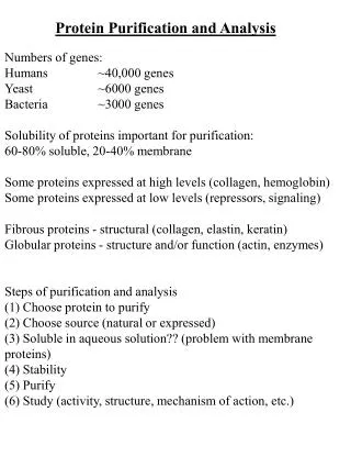
This detailed guide explores various techniques for purifying and characterizing proteins from cells, essential for biochemical research and industrial applications. It covers fundamental methods such as homogenization, differential centrifugation, salting in and out, dialysis, and various chromatography techniques including size-exclusion, affinity, and ion-exchange. Additionally, it discusses electrophoresis methods, immunoassays like ELISA, and protein sequencing techniques including Edman degradation and mass spectrometry. Each method's principles and applications are outlined for effective protein analysis and research.

Protein Purification and Characterization Techniques
E N D
Presentation Transcript
Protein Purificationand CharacterizationTechniques Nafith Abu Tarboush, DDS, MSc, PhD natarboush@ju.edu.jo www.facebook.com/natarboush
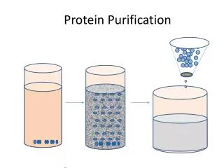
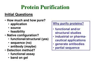
Extracting Pure Proteins from Cells • Purification techniques focus mainly on size & charge • The first step is homogenization (grinding, Potter–Elvejhem homogenizer, sonication, freezing and thawing, detergents) • Differential centrifugation (600 g: unbroken cells & nuclei; 15,000 g: mitochondria; 100,000 g: ribosomes and membrane fragments)
Salting in & out • Are proteins soluble? If yes, to which limit? • Salt stabilizes the various charged groups on a protein molecule and enhance the polarity of water, thus attracting protein into the solution and enhancing the solubility of protein • Ammonium sulfate is the most common reagent to use at this step • This technique is important but results are crude
Dialysis • Principle of diffusion • Concept of MW cut-off • Pure vs. crude
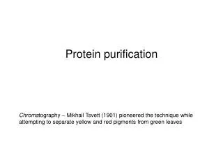
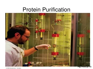
Column Chromatography • Greek chroma, “color,” and graphein, “to write” • Is it just for colourful proteins? • Chromatography is based on two phases: stationary & mobile • What are the different kinds?
Size-exclusion chromatographyGel-filtration chromatography • Separation on the basis of size (MW) • Stationary (cross-linked gel particles): consist of one of two kinds of polymers; the 1st is a carb. polymer (ex. dextran or agarose); often referred to by Sephadex and Sepharose. The 2nd is based on polyacrylamide (Bio-Gel) • Extent of crosslinking & pore size (exclusion limit) • Convenient & MW estimate • Each gel has range of sizes that separate linearly with the log of the molecular weight
Affinity chromatography • It has specific binding properties • The polymer (stationary) is covalently linked to a ligand that binds specifically to the desired protein • The bound protein can be eluted by adding high conc. of the soluble ligand • Protein–ligand interaction can also be disrupted with a change in pH or ionic strength • Convenient & products are very pure (Antigen-antibody, His-tag, GST-Tag)
Ion-exchange chromatography • Interaction based on net charge & is less specific • Resin is either negatively charged (cation exchanger) or positively charged (anion exchanger) • Buffer equilibration, exchange resin is bound to counter-ions. A cation-exchange resin is usually bound to Na+ or K+ ions, and an anion exchanger is usually bound to Cl– ions • Proteins mixture loading • Elution (higher salt concentration)
Electrophoresis • Based on the motion of charged particles in an electric field • Macromolecules have differing mobilities based on their charge, shape, and size • The most common medium is a polymer of agarose or acrylamide
Agarose vs. PAGE • Agarose (nucleic acids), PAGE (proteins) • In PAGE: SDS or NO-SDS {CH3(CH2)10CH2OSO3Na+} • SDS completely denatures proteins (multi-subunit proteins) • Acrylamide offers higher resistance to large molecules • Shape and charge are approximately the same (sizes is the determining factor) • Acrylamide without the SDS (native gel): study proteins in their native conformation (mobility is not an indication of size)
Isoelectric focusing • Proteins have different isoelectric points • Gel prepared with a pH gradient parallel to electric-field gradient • Two-dimensional gel electrophoresis (2-D gels)
Immunoassays – Western blot • From gel to a membrane (nitrocellulose or polyvinylidene difluoride, PVDF) • Detection: • Colorimetric: enzymes bound to 2nd Ab • Chemiluminescent: reporter 2nd Ab • Radioactive detection: X-rays • Fluorescent detection: fluorescently labeled probe
Immunoassays - ELISA • Enzyme-Linked Immunosorbent Assay • Detect & quantify substances ( peptides, proteins, antibodies & hormones) • Usually done in 96-well polystyrene plates (passively bind antibodies and proteins) • Apllication: • Screening (HIV, Hepatitis B&C) • Hormones (HCG, LH, TSH, T3, T4) (Green, positive) (No color, negative)
Protein sequencing - Edman Method • Step 1: how much and which amino acids are involved • Hydrolysis (heating + HCl) & Separation (ion-exchange chromatography or by high performance liquid chromatography, HPLC)
Protein sequencing - Edman Method • Step 2: determining the identities of N-terminal and C-terminal ends of protein • Necessary esp. to determine if the protein consists of one or two polypeptide chains • Steps 3: cleavage into smaller fragments (Edman degradation) • Enzymes- Trypsin, Chymotrypsin • Chemical reagents- Cyanogen bromide CNBr
Trypsin: Cleaves @ C-terminal of (+) charged side chains • Chymotrypsin: Cleaves @ C-terminal of aromatics • CNBr: Cleaves @ C-terminal of INTERNAL methionines
Protein sequencing – Mass Spectrometry • Mass/charge ratio
Protein sequencing – prediction from DNA & RNA • If the sequence of the gene is known, this is very easy • If the sequence of the gene is unknown (newly isolated proteins)? Sequence a short segment, complementary RNA, isolate mRNA, PCR, gene sequencing